Figures & data
Figure 1 A variety of bioabsorbable implants for use in spine applications.
Notes: Copyright © 2004 American Association of Neurological Surgeons. Reprinted with permission from the JNS Publishing Group. Reprinted from: Robbins MM, Vaccaro AR, Madigan L. The use of variables affecting convection implants in spine surgery. Neurosurg Focus. 2004;16(3):E1. http://thejns.org/.Citation205
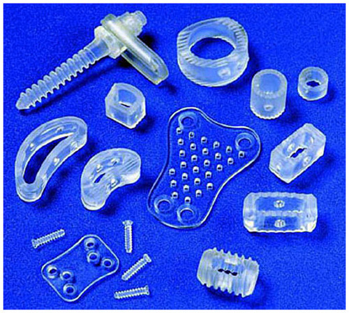
Figure 2 Tissue engineering and regenerative medicine rely on the implementation of various cell-, biomolecule-, and scaffold-based approaches to restore structure and function to developing and/or damaged tissues.
Note: Factors can be applied individually or in combination to achieve their desired effects.Citation206
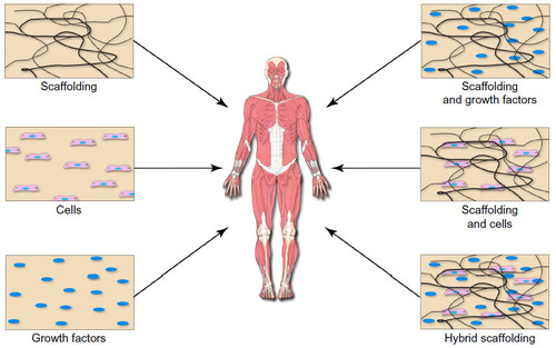
Figure 3 Characteristics of vancomycin-loaded chitosan coating.
Notes: (A) The total amount of drug loaded in the chitosan matrix during fabrication was determined as a function of vancomycin concentration in the suspension. (B) At the early stage, a burst release of vancomycin from the composite coatings was noticed. Afterwards, almost no release was detected for a certain period (approximately 90 hours). At the late stage, gradual release of the glycopeptides antibiotic was observed. (C) Accumulative weight loss of the coatings in PBS solution over time. The amount of chitosan weight loss was relatively noticeable after a long period of incubation (90 hours). (D) Surface morphology of chitosan coating after 7 day incubation in the PBS solution at 37°C. The presence of many pores revealed slow degradation and detachment of the coating in PBS. Reprinted from Mater Sci Eng C Mater Biol Appl, 41, Ordikhani F, Tamjid E, Simchi A, Characterization and antibacterial performance of electrodeposited chitosan-vancomycin composite coatings for prevention of implant-associated infections, 240–248, Copyright © 2014, with permission from Elsevier.Citation170
Abbreviations: EPD, electrophoretic deposition; PBS, phosphate-buffered saline.
Abbreviations: EPD, electrophoretic deposition; PBS, phosphate-buffered saline.
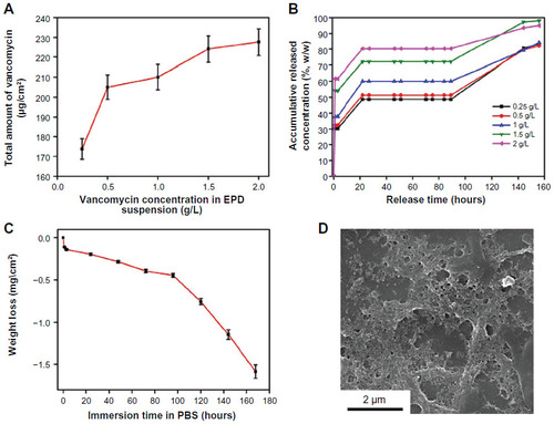
Figure 4 Scanning electron microscope images of a bioactive glass coating at different time-points.
Notes: The approximate thickness of the fabricated bioactive glass coating was 10 μm (A). The coating almost retained its thickness after 2 and 4 weeks of implantation (B and C). But, the images also indicate dissolution of the coating after the healing periods. The EDS analysis (D–F) showed the presence of Ca and P (ie, consistent with calcium phosphate) in the coating even after 4 weeks of implantation. D–F are the EDS spectra corresponding to A (before implantation), B (2 weeks after implantation), and C (4 weeks after implantation), respectively. Reproduced from Chaudhari A, Braem A, Vleugels J, et al. Bone tissue response to porous and functionalized titanium and silica based coatings. PLoS One. 2011;6(9):e24186.Citation173
Abbreviations: Acc, accelerating; BAG, bioactive glass; BSE, backscatter electrons; Det, detector; EDS, energy-dispersive X-ray spectroscopy; Magn, magnification; WD, working distance.
Abbreviations: Acc, accelerating; BAG, bioactive glass; BSE, backscatter electrons; Det, detector; EDS, energy-dispersive X-ray spectroscopy; Magn, magnification; WD, working distance.
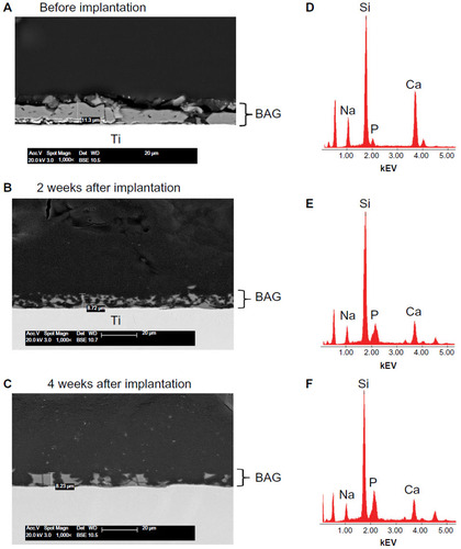
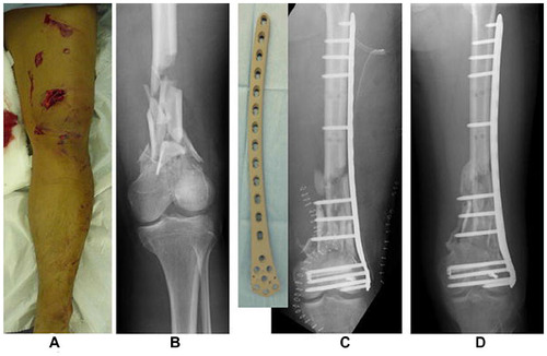
Figure 5 Case of a 42 year-old patient who was treated with an iodine-coated internal fixation plate.
Notes: A Gustilo grade IIIa open fracture (A: photograph; B: X-ray) treated with an iodine-supported titanium plate (C). Bony union was established with good callus formation at 4 months postsurgery, with no signs of infection (D). Reproduced from Tsuchiya H, Shirai T, Nishida H, et al. Innovative antimicrobial coating of titanium implants with iodine. J Orthop Sci. 2012;17(5):595–604.Citation183