
Figures & data
Figure 1 Tissue microarray and immunohistochemistry staining of P-cofilin in invasive ductal breast cancer tumors.
Abbreviation: P-cofilin, phosphorylated cofilin.
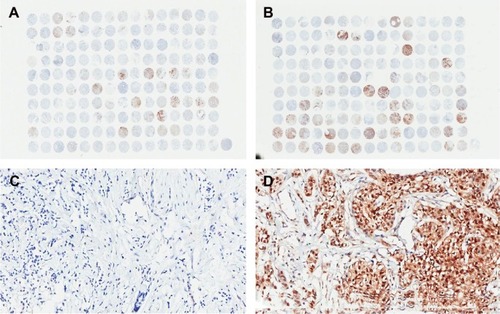
Figure 2 Association of P-cofilin with overall survival among patients with invasive ductal breast cancer.
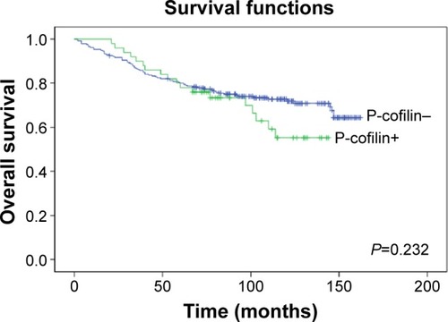
Figure 3 Association of cofilin with overall survival among P-cofilin-negative patients with invasive ductal breast cancer.
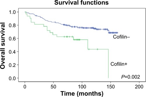
Figure 4 Association of cofilin with overall survival among P-cofilin-positive patients with invasive ductal breast cancer.
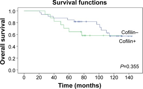
Table 1 Cofilin with or without phosphorylation for predicting overall survival in invasive ductal breast cancer
Table 2 Clinicopathological characteristics of breast cancer with different cofilin expression
Table 3 Univariate and multivariate analysis of factors that predict overall survival in invasive ductal breast cancer