
Figures & data
Figure 1 Computed tomography scans showing a homogeneous, isolated, ill-defined soft tissue density mass in the right orbit (A–C; arrows). The right eyeball was displaced anteriorly and inferiorly. (A) Axial section, (B) sagittal section, and (C) coronal section.
Abbreviations: AR, anterior; L, left; PI, posterior; R, right.
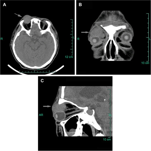
Figure 2 Histopathological sections of the mass.
Notes: (A) Irregularly shaped nests of tumor cells surrounded by avascularized fibrous stroma (arrow shows the avascularized fibrous stroma; hematoxylin and eosin staining; original magnification: ×100). (B) The monomorphic malignant tumor cells are small round blue cells that are slightly larger than mature lymphocytes, with a very high nuclear to cytoplasmic ratio (hematoxylin and eosin staining; original magnification: ×400).
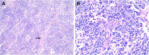
Figure 3 Immunohistochemical staining of the mass.
Notes: The tumor cells were positive for (A) SYN; original magnification: ×400; a reaction in the cytoplasm of the neoplastic cells; (B) CGA; original magnification: ×400; a reaction in the cytoplasm of the neoplastic cells; (C) CD56 (cluster of differentiation 56); original magnification: ×400; a strong and heavy membrane-type staining; (D) CK; original magnification: ×400; a reaction in the cytoplasm of the neoplastic cells; (E) VIM; original magnification: ×400; a reaction in the cytoplasm of the neoplastic cells; (F) S-100 protein; original magnification: ×400; a sustentacular reaction. The tumor cells were negative for EMA and CD99 (data not shown).
Abbreviations: CD, cluster of differentiation; CGA, chromogranin; CK, cytokeratin; EMA, epithelial membrane antigen; SYN, synaptophysin; VIM, vimentin.
Abbreviations: CD, cluster of differentiation; CGA, chromogranin; CK, cytokeratin; EMA, epithelial membrane antigen; SYN, synaptophysin; VIM, vimentin.
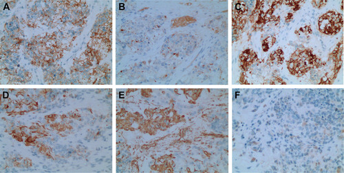
Table 1 Selected immunohistochemical results for malignant small round cell tumors