Figures & data
Figure 1 The anticancer activities of EGCG against HuCC-T1 cells.
Notes: (A) The effects of EGCG on the viability of cancer cells. A total of 3×10⁴ cells were used to measure the anticancer effect and 3×10³ cells were used to test growth inhibition. RPMI1640 media supplemented with 10% FBS was used to assess tumor cell growth inhibition and serum-free media was used to assess the anti-cancer effects. (B) TUNEL staining. Images were observed at 400×. (Numbers in the boxes indicate intensities of FITC.) (C) Flow cytometric analysis of cancer cells. FITC Annexin V and PI were used for apoptosis and necrosis analysis of tumor cells, respectively. *,**P<0.01.
Abbreviations: IC50, half maximal inhibitory concentration; RPMI1640, Roswell Park Memorial Institute 1640; EGCG, epigallocatechin-3-gallate; FBS, fetal bovine serum; FITC, fluorescein isothiocyanate; HuCC-T1, human cholangiocellular carcinoma cell line; HEK 293T, human embryonic kidney 293 T; PI, propidium iodide; TUNEL, terminal deoxynucleotidyl transferase dUTP nick end labeling.
Abbreviations: IC50, half maximal inhibitory concentration; RPMI1640, Roswell Park Memorial Institute 1640; EGCG, epigallocatechin-3-gallate; FBS, fetal bovine serum; FITC, fluorescein isothiocyanate; HuCC-T1, human cholangiocellular carcinoma cell line; HEK 293T, human embryonic kidney 293 T; PI, propidium iodide; TUNEL, terminal deoxynucleotidyl transferase dUTP nick end labeling.
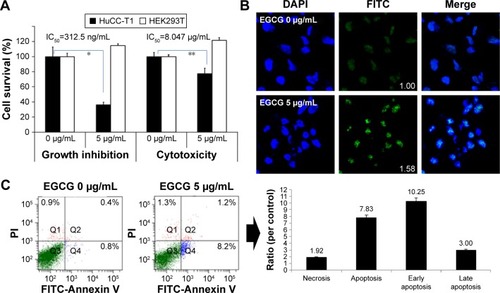
Figure 2 The apoptosis and necrosis of HuCC-T1 cells by treatment of EGCG.
Notes: Western blot assay: (A) wt-p53, mut-p53; (C) Bax, Bad, Bcl-2, Caspase-3, Caspase-9, and PARP expression. Fluorescence microscopic observation: (B) mut-p53 (Numbers in the boxes indicate intensities of green fluorescence.); (D) Bax, Bcl-2, Caspase-3, Caspase-9, and PARP expression. (E) The extent of Caspase-3,9 activities. Images were observed at 400×. *P<0.01.
Abbreviations: wt, wild-type; mut, mutant; EGCG, epigallocatechin-3-gallate; GAPDH, glyceraldehyde 3-phosphate dehydrogenase; HuCC-T1, human cholangiocellular carcinoma cell line, PARP, poly adenosine diphosphate ribose polymerase.
Abbreviations: wt, wild-type; mut, mutant; EGCG, epigallocatechin-3-gallate; GAPDH, glyceraldehyde 3-phosphate dehydrogenase; HuCC-T1, human cholangiocellular carcinoma cell line, PARP, poly adenosine diphosphate ribose polymerase.
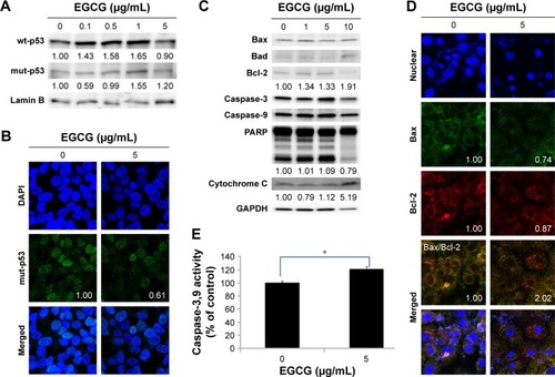
Figure 3 The effects of EGCG on the MMP activity, invasion and migration capacity of HuCC-T1 cells.
Notes: (A) Gelatin zymography: MMP-2 and -9 expressions. (B) Matrigel® invasion assay. (C) Wound healing assay for cancer cell migration. Images were observed at 400×. *P<0.001.
Abbreviations: EGCG, epigallocatechin-3-gallate; HuCC-T1, human cholangiocellular carcinoma cell line; MMP, matrix metalloproteinase.
Abbreviations: EGCG, epigallocatechin-3-gallate; HuCC-T1, human cholangiocellular carcinoma cell line; MMP, matrix metalloproteinase.
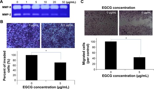
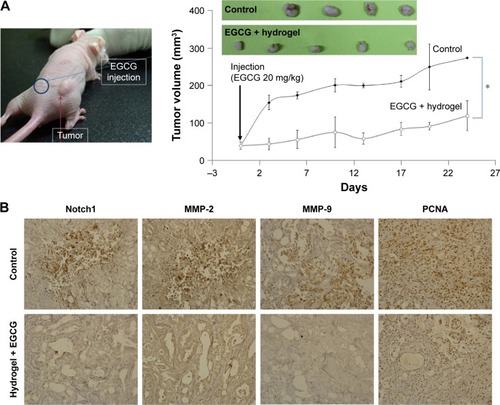
Figure 4 Antitumor activities of EGCG against HuCC-T1 tumor xenograft mice model.
Notes: (A) Tumor growth. 1×10⁷ cells were subcutaneously injected to the back of BALB/c nude mice. When the diameter of the solid tumor reached about 4 mm, EGCG in a vehicle (hydrogel) was injected subcutaneously beside the solid tumor (dose: 20 mg EGCG/kg). For comparison, the vehicle as a control was injected into the back of the mice. Growth of the tumor was calculated using the formula V = (a × [b]Citation2)/2, with a being the largest and b being the smallest diameter. For immunohistochemistry of tumor tissues, tumors were isolated and fixed with 4% formamide 25 days after the injection. (B) Immunohistochemistry (400×) of tumor tissues. Notch 1, MMP-2 and -9, and PCNA antibodies were used for staining tumor tissues. *P<0.01.
Abbreviations: EGCG, epigallocatechin-3-gallate; HuCC-T1, human cholangiocellular carcinoma cell line; MMP, matrix metalloproteinase; PCNA, proliferating cell nuclear antigen.
Abbreviations: EGCG, epigallocatechin-3-gallate; HuCC-T1, human cholangiocellular carcinoma cell line; MMP, matrix metalloproteinase; PCNA, proliferating cell nuclear antigen.