
Figures & data
Table 1 The clinical information of primary NEBC patients at diagnosis
Table 2 The immunohistochemistry of primary NEBC patients
Table 3 The adjuvant therapy and clinical follow-up of primary NEBC patients
Figure 1 CT image of the patient with hepatic metastasis in September 2012, 70 months after the initial surgical treatment.
Notes: (A and B) The image of plain scan showed that the density of liver parenchyma was diffusely reduced, and extensive round-like higher density lesions in the liver. (C and D) Enhanced CT revealed multiple nodular enhancement in liver.
Abbreviation: CT, computed tomography.
Abbreviation: CT, computed tomography.
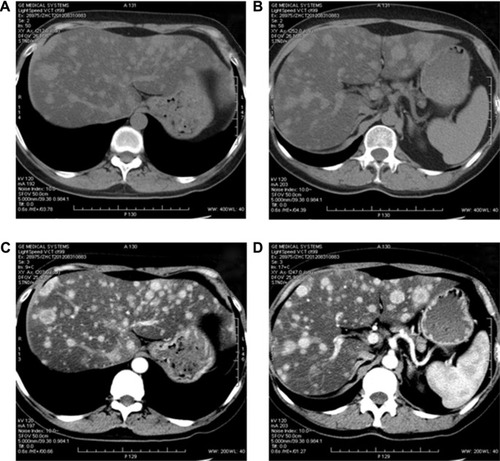
Figure 2 Post-therapy abdominal CT of the patient with hepatic metastasis in April 2013.
Note: Over a period of 7 months of treatment, both (A and B) plain scan and (C and D) enhanced CT showed that the hepatic lesions were gradually decreased.
Abbreviation: CT, computed tomography.
Abbreviation: CT, computed tomography.
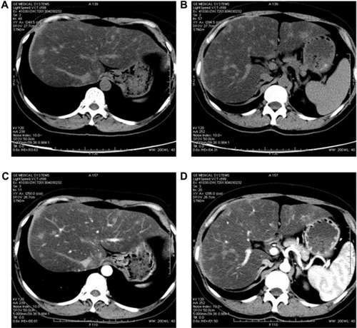
Figure 3 Post-therapy abdominal CT of the patient with hepatic metastasis in May 2014.
Note: With 20 months of treatment, the hepatic lesions were further reduced in both (A and B) plain scan and (C and D) enhanced CT.
Abbreviation: CT, computed tomography.
Abbreviation: CT, computed tomography.
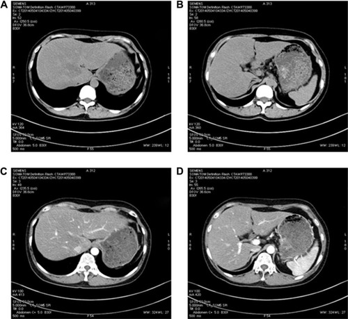
Figure 4 Surveillant CT.
Notes: (A and B) The CT of the abdomen showed that the hepatic metastasis lesions of this patient progressed again in October 2014. (C and D) After a modified therapy, the reduction of hepatic lesions was obviously found by CT of the abdomen in February 2015.
Abbreviation: CT, computed tomography.
Abbreviation: CT, computed tomography.
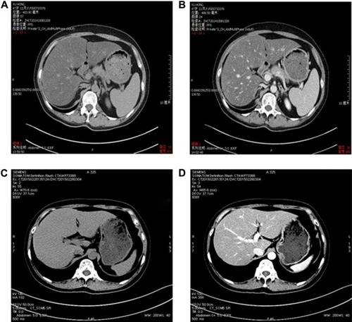
Table S1 Mammography features of NEBC
Table S2 Ultrasonography features of NEBC