
Figures & data
Figure 1 Lymphoscintigraphy including the area from the hand to the abdominal region.
Source: Reprinted with permission from Sarri AJ, Moriguchi SM, Dias R, et al. Physiotherapeutic stimulation: Early prevention of lymphedema following axillary lymph node dissection for breast cancer treatment. Exp Ther Med. 2010;1(1): 147–152.Citation29
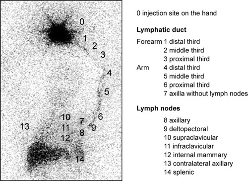
Table 1 Descriptive statistics of the sample ALND and SLNB groups
Table 2 Types of surgery in ALND and SLNB groups
Table 3 Sites reached by the lymphatic flow in the ALND and SLNB groups distributed over the dynamic, static, and WBS image stages and classified into forearm, arm/axilla, and thoracic extra-axillary lymph nodes
Figure 2 Difference in lymphatic progression between the (A) ALND and (B) SLNB groups. WBS of the ALND and SLNB groups.
Abbreviations: ALND, axillary lymph node dissection; SLNB, sentinel lymph node biopsy; WBS, whole-body scan.
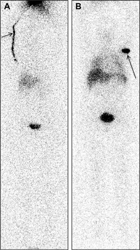
Figure 3 WBS of the ALND group.
Abbreviations: ALND, axillary lymph node dissection; WBS, whole-body scan.
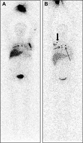
Figure 4 Lymphoscintigraphy. Different distributions of lymph nodes in the ALND and SLNB groups.
Abbreviations: ALND, axillary lymph node dissection; SLNB, sentinel lymph node biopsy; WBS, whole-body scan.
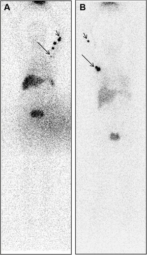
Table 4 Location of lymph nodes in the ALND and SLNB groups observed in the dynamic, static, and WBS imaging