Figures & data
Table 1 Association between CUL4A expression and clinicopathologic features of colorectal cancer patients
Figure 1 CUL4A is overexpressed and associated with prognosis in colorectal cancer.
Notes: (A) The expression of CUL4A was detected in colorectal normal tissue and cancer tissue by immunohistochemistry, and representative samples are shown at 400× magnification. (B) Comparison of mRNA expression levels of CUL4A in 10 pairs of tumor and adjacent normal samples with qRT-PCR. (C) The overall survival rate of the 80 colorectal cancer patients were compared in the CUL4A low and CUL4A high groups. Statistical significance was determined using the log-rank test.
Abbreviation: qRT-PCR, quantitative reverse transcription-polymerase chain reaction.
Abbreviation: qRT-PCR, quantitative reverse transcription-polymerase chain reaction.
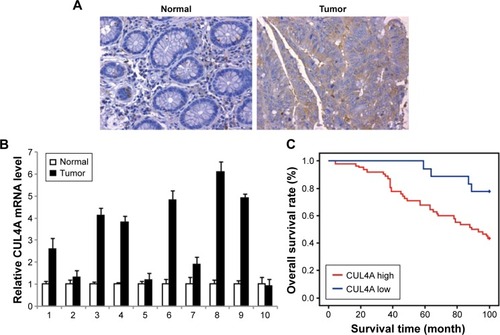
Figure 2 CUL4A promotes colorectal cancer cell line HCT-116 growth and migration.
Notes: (A) The mRNA level of CUL4A in HCT-116 cells infected with lentivirus containing vector or CUL4A was detected by qRT-PCR. (B) The mRNA level of CUL4A in HCT-116 cells infected with lentivirus containing mock or shCUL4A was detected by qRT-PCR. (C) Cell proliferation rate of vector or CUL4A cells were determined by MTT assay at the indicated time point. (D) Cell proliferation rate of mock or shCUL4A cells were determined by MTT assay at the indicated time point. The statistical results are shown in the lower panel. Effect of CUL4A overexpression (E) and knockdown (F) on colony formation were measured in HCT-116 cells. The cells were seeded into 6-well plates and cultured for 5 days, followed by crystal violet staining. The colony counts were shown below the graph. All the data are shown as mean ± standard deviation and *P≤0.05, **P≤0.01, ***P≤0.001.
Abbreviations: MTT, 3-(4,5-dimethylthiazol-2-yl)-2,5-diphenyltetrazolium bromide; qRT-PCR, quantitative reverse transcription-polymerase chain reaction.
Abbreviations: MTT, 3-(4,5-dimethylthiazol-2-yl)-2,5-diphenyltetrazolium bromide; qRT-PCR, quantitative reverse transcription-polymerase chain reaction.
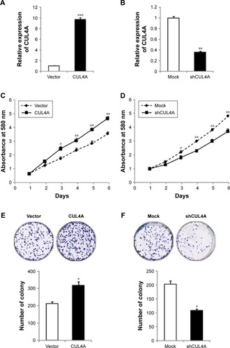
Figure 3 CUL4A promotes migration of colorectal cancer cell line HCT-116.
Notes: (A and B) CUL4A overexpressing and knockdown HCT-116 cells or control vector cells were subjected to wound healing assay. The statistical results are shown in the lower panel. Data are shown as mean ± SD. *P≤0.05, **P≤0.01.
Abbreviations: SD, standard deviation; h, hours.
Abbreviations: SD, standard deviation; h, hours.
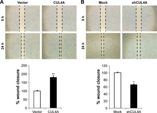
Figure 4 CUL4A promotes epithelial–mesenchymal transition by changing the H3K4me3 level in promoter regions of E-cadherin, N-cadherin and vimentin.
Notes: (A and B) Expression of epithelial and mesenchymal markers was analyzed by Western blotting and qRT-PCR. (C) Quantitative chromatin immunoprecipitation was performed to assess H3K4me3 occupancy in HCT-116 CUL4A overexpression cells. IgG was used as the negative control. “% input” indicates the ratio of DNA fragment of each promoter region bound by H3K4me3 to the total amount of input DNA fragment without H3K4me3 antibody pull-down. All the data are shown as mean ± standard deviation and *P≤0.05, **P≤0.01.
Abbreviations: GAPDH, glyceraldehyde 3-phosphate dehydrogenase; IgG, immunoglobulin G; H3K4me3, H3K4 trimethylation; PCR, polymerase chain reaction; qRT-PCR, quantitative reverse transcription-polymerase chain reaction.
Abbreviations: GAPDH, glyceraldehyde 3-phosphate dehydrogenase; IgG, immunoglobulin G; H3K4me3, H3K4 trimethylation; PCR, polymerase chain reaction; qRT-PCR, quantitative reverse transcription-polymerase chain reaction.
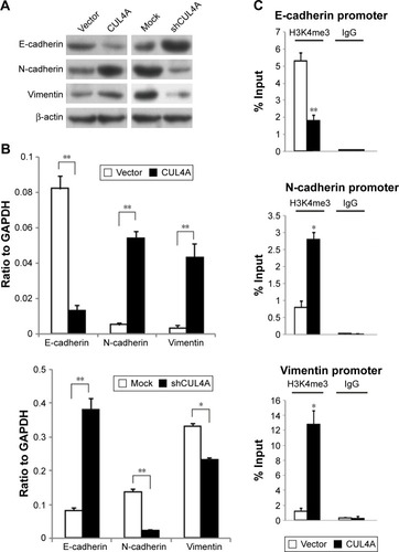
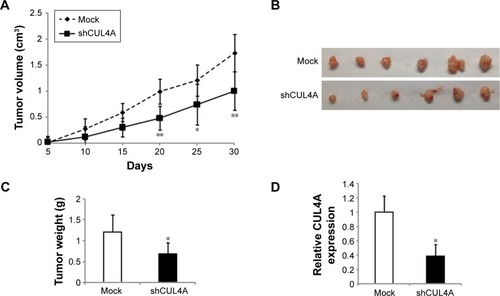
Figure 5 Knockdown of CUL4A suppresses tumor growth in vivo.
Notes: (A and B) Tumor volume was calculated every 5 days after the injection of HCT-116 cells with knockdown of CUL4A. Error bars indicate SD. (C) Tumor weight is represented as mean of tumor weights ±SD. (D) qPCR analysis of CUL4A expression in tumor tissues formed from HCT-116/mock and HCT-116/shCUL4A. *P≤0.05, **P≤0.01.
Abbreviations: qPCR, quantitative polymerase chain reaction; SD, standard deviation.
Abbreviations: qPCR, quantitative polymerase chain reaction; SD, standard deviation.