Figures & data
Table 1 The primers used for real-time PCR
Figure 1 Isoorientin inhibits cell proliferation.
Notes: Two cell lines were treated with isoorientin at the concentrations of 0, 20, 40, 80, and 160 μM for 24 hours. (A) The fold change in cell viability of PATU-8988 and PANC-1 cells. (B) The fold change in cell viability of shRNA–PATU-8988 and shRNA–PANC-1 cells. The data represent the mean ± SEM (n=3, *P<0.05 vs 0 μM group).
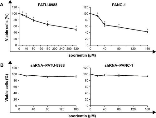
Figure 2 AMPK is constitutively activated by isoorientin.
Notes: The effect of isoorientin on the expression of the protein 5-AMP-activated protein kinase (AMPK). (A) The expression levels of AMPK and phosphorylated AMPK were evaluated in PATU-8988 and PANC-1 cell lines in the absence (−) or presence (+) of isoorientin (20, 40, 80, and 160 μM) for 24 hours. GAPDH was used as a loading control. (B) The expression of AMPK and p-AMPK in the shRNA–PATU-8988 and shRNA–PANC-1 PC groups. The immunoblots shown are representative of three independent experiments. NC, transfected with negative control lentivirus PC cell group. shRNA, shRNA-interfering PC group. WT, wild-type PC cell.
Abbreviations: AMPK, AMP-activated protein kinase; PC, pancreatic cancer.
Abbreviations: AMPK, AMP-activated protein kinase; PC, pancreatic cancer.
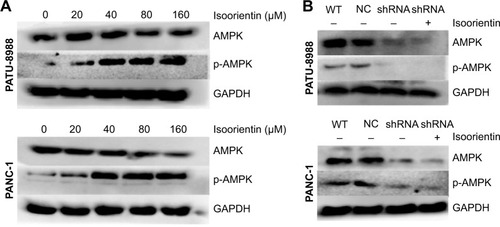
Figure 3 Pancreatic cancer cells are susceptible to isoorientin-induced apoptosis.
Notes: The effect of isoorientin on apoptotic cell death. Cells were treated in the absence or presence of isoorientin (20, 40, 80, and 160 μM) for 24 hours. Apoptosis was evaluated by Annexin V staining (A, B, D, E). The expression of BCL-2 and BAX was evaluated by Western blotting(C and F). GAPDH was used as a loading control. The immunoblots shown are representative of three independent experiments. The apoptosis data are presented as the mean ± SEM from three independent experiments (*P<0.05 vs 0 μM).
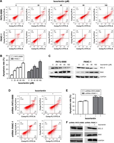
Figure 4 Isoorientin downregulates the expression of VEGF, MMPs, and proteins involved in the epithelial–mesenchymal transition.
Notes: PATU-8988 and PANC-1 cells were treated with isoorientin at the concentrations of 0, 20, 40, 80, and 160 μM for 24 hours. (A) The fold change in the mRNA expression of CDH1 (E-cadherin), CDH2 (N-cadherin), MMP2, MMP9, and VEGF of the two cell lines. (B) A Western blotting assay was performed to determine the protein levels of E-cadherin, N-cadherin, MMP2, MMP9, and VEGF. GAPDH was used as an internal control. (E) The releasing VEGF level in medium test by ELISA. (C–E) The fold change in the expression of CDH1 (E-cadherin), CDH2 (N-cadherin), MMP2, MMP9, and VEGF in shRNA–PATU-8988 and shRNA–PANC-1 cells with (+) or without (−) isoorientin treatment was quantified by qRT-PCR, Western blotting, and ELISA. The data represent the mean ± SEM (n=3, *P<0.05 vs 0 μM group).
Abbreviation: ELISA, enzyme-linked immunosorbent assay; MMP, matrix metalloproteinases; qRT-PCR, quantitative real-time PCR; VEGF, vascular endothelial growth factor.
Abbreviation: ELISA, enzyme-linked immunosorbent assay; MMP, matrix metalloproteinases; qRT-PCR, quantitative real-time PCR; VEGF, vascular endothelial growth factor.
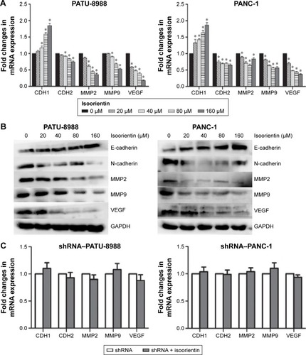
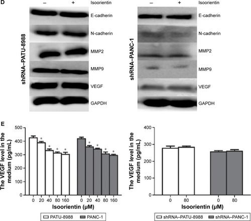
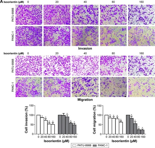
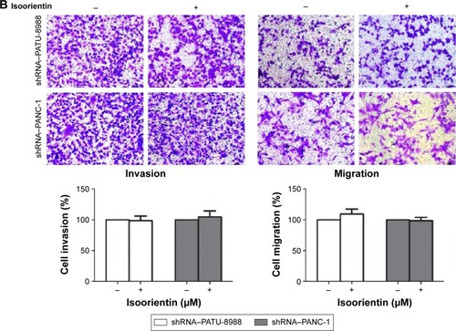
Figure 5 Isoorientin inhibits the migration and invasion of pancreatic cancer cells.
Notes: Two cell lines were treated with luteolin at the concentrations of 0, 20, 40, 80, and 160 μM for 24 hours. (A) Isoorientin inhibited the invasive and migration capacity of PATU-8988 and PANC-1 cells. 200× magnification. (B) In the shRNA–PATU-8988 and shRNA–PANC-1 cell lines, isoorientin had little effect on their invasive and migration capacity. The data represent the mean ± SEM (n=3, *P<0.05 vs 0 μM group). 200× magnification.