Figures & data
Table 1 Primer sequences used for qRT-PCR
Figure 1 miR-320a relative expression in HCC samples and cell lines.
Notes: (A) miR-320a levels were accessed by qRT-PCR in HCCs and their adjacent nontumor tissues. Small nuclear RNA U6 was used as an internal control. (B) The expression of miR-320a was measured in five normal liver tissue, one normal cell line and four HCC cell lines by qRT-PCR, and the expression levels of miR-320a were normalized to U6. Data are presented as the mean ± standard deviation (n=3). Results were obtained in three replicates. *P<0.05.
Abbreviations: HCC, hepatocellular carcinoma; miR-320a, microRNA-320a; qRT-PCR, quantitative reverse-transcription polymerase chain reaction; ns, nonsignificant.
Abbreviations: HCC, hepatocellular carcinoma; miR-320a, microRNA-320a; qRT-PCR, quantitative reverse-transcription polymerase chain reaction; ns, nonsignificant.
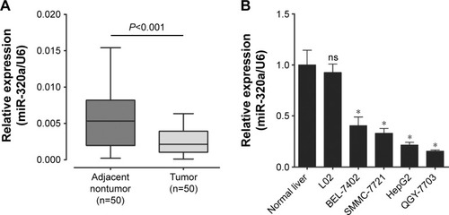
Figure 2 miR-320a directly binds to the coding DNA sequence of c-Myc.
Notes: (A) Schematic representation of the c-Myc messenger RNA with two putative miR-320a-binding sites in the CDS. (B) Schematic representation of luciferase reporter constructs. Two sequences including A or B sites were cloned into the luciferase reporter plasmid individually or in combination. (C) Luciferase reporter assay to verify the activity of miR-320a upon the target site of c-Myc. HEK-293 cells were transfected with luciferase reporter plasmid, miR-320a mimics, or NC. The firefly luciferase activity was normalized to the Renilla luciferase activity, and the normalized luciferase activity of the negative control was set as relative luciferase activity 1. Data are shown as mean ± standard deviation (n=3). Results were obtained in three replicates. *P<0.05, **P<0.01.
Abbreviations: CDS, coding DNA sequence; DNA, deoxyribonucleic acid; NC, negative control; miR-320a, microRNA-320a; UTR, untranslated regions.
Abbreviations: CDS, coding DNA sequence; DNA, deoxyribonucleic acid; NC, negative control; miR-320a, microRNA-320a; UTR, untranslated regions.
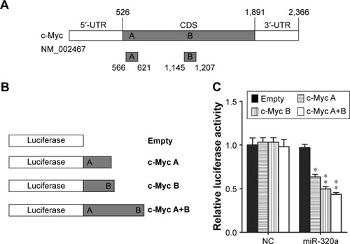
Figure 3 miR-320a downregulates endogenous c-Myc expression in HCC cells.
Notes: (A) The upregulation of miR-320a was verified by qRT-PCR after transfected with miR-320a mimics or NC in HepG2 and QGY-7703 cells. U6 was used as an internal control. (B) The protein levels of c-Myc expression were determined by Western blot after transfected with miR-320a mimics or NC in HepG2 and QGY-7703 cells. β-Actin was used as a control. (C) Analysis of c-Myc and miR-320a expression in the same HCC tissue by IHC and qRT-PCR. Brown signal in IHC was considered as positive staining for c-Myc. Scale bar =50 μm. Data are presented as the mean ± standard deviation (n=3). Results were obtained in three replicates. *P<0.05, **P<0.01.
Abbreviations: HCC, hepatocellular carcinoma; IHC, immunohistochemistry; miR-320a, microRNA-320a; NC, negative control; qRT-PCR, quantitative reverse-transcription polymerase chain reaction.
Abbreviations: HCC, hepatocellular carcinoma; IHC, immunohistochemistry; miR-320a, microRNA-320a; NC, negative control; qRT-PCR, quantitative reverse-transcription polymerase chain reaction.
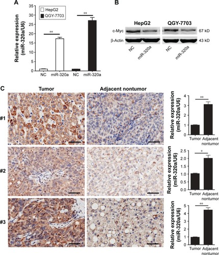
Figure 4 Upregulation of miR-320a inhibited c-Myc expression and hepatocellular carcinoma cells proliferation by targeting c-Myc in vitro.
Notes: After HepG2 and QGY-7703 cell lines treated with miR-320a mimics or c-Myc siRNA. Western blot assays of c-Myc protein were conducted in HepG2 (A) and QGY-7703 (B) cells and β-Actin was used as a control. 3-(4,5-Dimethylthiazol-2-yl)-2,5-diphenyl tetrazolium bromide assays were conducted in HepG2 cell line (C) and QGY-7703 cell line (D). Data are presented as the mean ± standard deviation (n=3). Results were obtained in three replicates. **P<0.01.
Abbreviations: miR-320a, microRNA-320a; NC, negative control; OD, optical density; si or siRNA, small interfering RNA.
Abbreviations: miR-320a, microRNA-320a; NC, negative control; OD, optical density; si or siRNA, small interfering RNA.
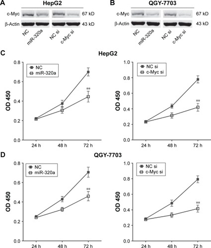
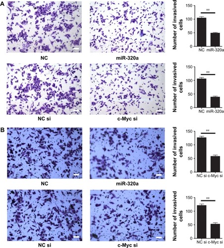
Figure 5 Upregulation of miR-320a inhibited hepatocellular carcinoma cells invasion by targeting c-Myc in vitro.
Notes: Transwell invasion assay was performed to detect the invasive properties in HepG2 (A) and QGY-7703 (B) cells after treated with miR-320a mimics or c-Myc siRNA; Scale bar =50 μm. Data are presented as the mean ± standard deviation (n=3). Results were obtained in three replicates. **P<0.01.
Abbreviations: miR-320a, microRNA-320a; NC, negative control; si or siRNA, small interfering RNA.
Abbreviations: miR-320a, microRNA-320a; NC, negative control; si or siRNA, small interfering RNA.