Figures & data
Table 1 Primer sequences
Figure 1 Expression pattern of WWC3 protein in gastric cancer tissues.
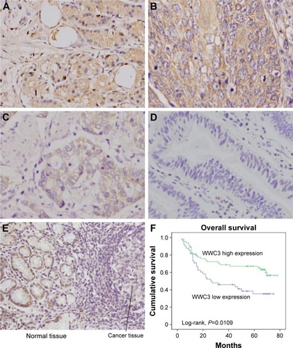
Table 2 Distribution of WWC3 status in gastric cancer according to clinicopathological characteristics
Table 3 Univariate and multivariate analyses for predictive factors in patients with gastric carcinoma (Cox regression model)
Figure 2 WWC3 inhibits gastric cancer cell proliferation.
Abbreviations: siRNA, small interfering RNA; PCR, polymerase chain reaction.
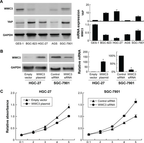
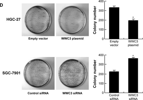
Figure 3 WWC3 regulates gastric cancer cell invasion and cell cycle progression.
Abbreviation: siRNA, small interfering RNA.
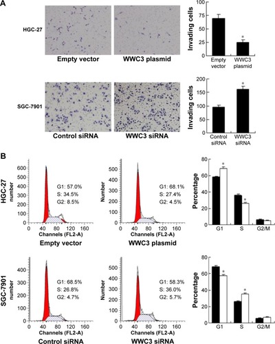
Figure 4 WWC3 regulates cell cycle-related proteins and Hippo signaling pathway.
Abbreviation: PCR, polymerase chain reaction.
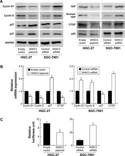
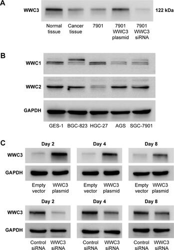
Figure S1 Validation of antibody and transfection efficiency.
Notes: (A) The specificity of the antibody was validated by using gastric cancer tissue, corresponding normal tissue, and cell lines with WWC3 overexpression and siRNA depletion. Western blot was used to examine WWC3 with this antibody. The Western blot bands of cells were strong when WWC3 was overexpressed and were weak when depleted. Gastric cancer tissues and normal tissues showed similar single bands at the same molecular weight. (B) Expression of WWC1 and WWC2 proteins in gastric cancer cell lines and GES-1 cell line. (C) The knockdown and overexpression efficiency were checked at different time points (at 2, 4, and 8 days after transfection) using Western blot. The transfection of plasmid significant upregulated WWC3 protein at day 8. The knockdown efficiency was most significant at day 2 and could still be observed at day 8.
Abbreviation: siRNA, small interfering RNA.