Figures & data
Figure 1 Expression of AIM2 mRNA and protein measured by RT-PCR and Western blotting.
Notes: (A) The expression of AIM2 mRNA dramatically increased after lentivirus-AIM2 transfection. (B) Western blotting analysis of AIM2 protein expression in NC and AE cells. (C) Relative expression of AIM2 was significantly upregulated in the AE group. **P<0.01, ***P<0.001.
Abbreviations: AE, AIM2-expressing group; AIM2, absent in melanoma 2; NC, negative control; RT-PCR, real-time polymerase chain reaction.
Abbreviations: AE, AIM2-expressing group; AIM2, absent in melanoma 2; NC, negative control; RT-PCR, real-time polymerase chain reaction.
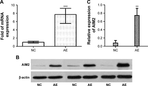
Figure 2 The effect of AIM2 on viability and apoptosis of HCT116 cells.
Notes: (A) MTT assay showed that the expression of AIM2 suppressed the cell viability compared with the negative control. (B) Cell apoptosis detected by Annexin V-APC dual-label flow cytometry. The apoptosis rate was significantly increased in the AE group; IGF-1 decreased the apoptosis rate of the AE group but had no significant effect in the NC group. **P<0.01; ***P<0.001.
Abbreviations: AE, AIM2-expressing group; AIM2, absent in melanoma 2; APC, Allophycocyanin; IGF-1, insulin-like growth factor-1; MTT, 3-(4,5-dimethylthiazol-2-yl)-2,5-diphenyltetrazolium bromide; NC, negative control; OD, optical density.
Abbreviations: AE, AIM2-expressing group; AIM2, absent in melanoma 2; APC, Allophycocyanin; IGF-1, insulin-like growth factor-1; MTT, 3-(4,5-dimethylthiazol-2-yl)-2,5-diphenyltetrazolium bromide; NC, negative control; OD, optical density.
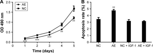
Figure 3 Cell cycle distribution of HCT116 CRC cells.
Notes: (A) NC and AE cells were fixed in ethanol, stained with PI, and analyzed by flow cytometry. (B) Expression of AIM2 increased the proportion of cells in S phase and decreased the proportion of cells in G1 and G2/M phases compared to control (n=3, *P<0.05, **P<0.01).
Abbreviations: AE, AIM2-expressing group; AIM2, absent in melanoma 2; CRC, colorectal cancer; NC, negative control; PI, propidium iodide.
Abbreviations: AE, AIM2-expressing group; AIM2, absent in melanoma 2; CRC, colorectal cancer; NC, negative control; PI, propidium iodide.
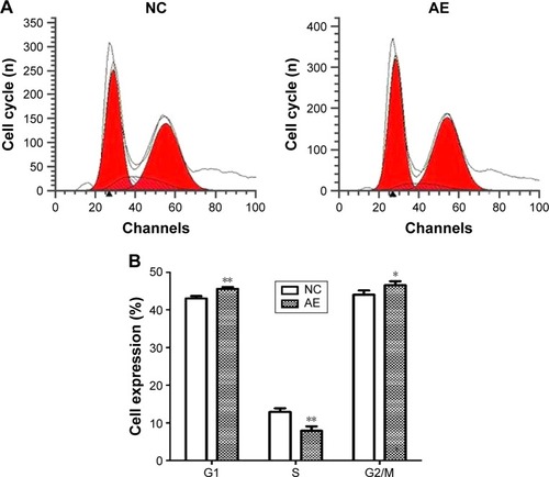
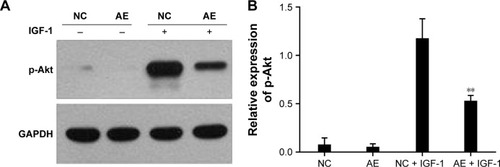
Figure 4 Effect of AIM2 expression on activation of the PI3K/Akt pathway in HCT116 cells.
Notes: (A) Western blot analysis of p-Akt protein level in NC and AE cells with and without IGF-1 treatment. (B) Quantification of the immunoreactivity showed no significant difference in the expression of p-Akt between NC and AE cells in the absence of IGF-1 (P=0.54), whereas after stimulation with IGF-1 the level of p-Akt was significantly lower in AE cells compared with NC cells (**P<0.01).
Abbreviations: AE, AIM2-expressing group; AIM2, absent in melanoma 2; Akt, protein kinase B; GAPDH, glyceraldehyde-3-phosphate dehydrogenase; IGF-1, insulin-like growth factor-1; NC, negative control; PI3K, phosphatidylinositol 3-kinase.
Abbreviations: AE, AIM2-expressing group; AIM2, absent in melanoma 2; Akt, protein kinase B; GAPDH, glyceraldehyde-3-phosphate dehydrogenase; IGF-1, insulin-like growth factor-1; NC, negative control; PI3K, phosphatidylinositol 3-kinase.