Figures & data
Figure 1 Expression of SIX1 protein in esophageal squamous cell carcinoma (ESCC) tissues.
Notes: (A) Negative SIX1 expression in normal esophageal squamous mucosa tissues. (B) Negative SIX1 expression in a case of stage 1, grade 1 ESCC tissue. (C) Positive nuclear staining of SIX1 in a case of grade 2 ESCC. (D) Positive nuclear and cytoplasmic expression in a case of stage 3 ESCC tissue. (E) Survival analysis of SIX1 in ESCC tissues. Magnification, 400×.
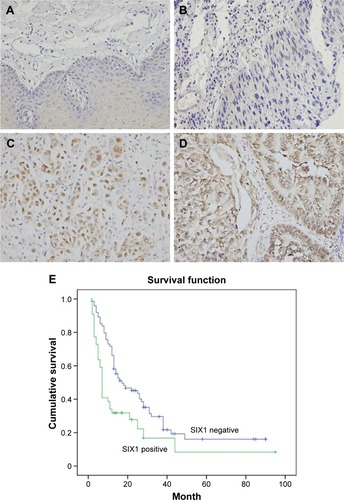
Table 1 Distribution of SIX1 status in esophageal carcinoma according to clinicopathological characteristics
Table 2 Multivariate analysis for predictive factors in patients with esophageal carcinoma (Cox regression model)
Figure 2 Expression of SIX1 in esophageal squamous cell carcinoma cell lines and its transfection and knockdown efficiency.
Notes: (A) Western blot showed that SIX1 protein was high in Eca-109 cell line and low in TE-1 cell line. (B) Western blot and real-time PCR analyses showed that SIX1 transfection in TE-1 cell line increased its protein and mRNA expression. SIX1 siRNA treatment in Eca-109 cell line decreased its protein and mRNA expression.
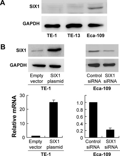
Figure 3 SIX1 promotes proliferation and invasion in esophageal squamous cell carcinoma cell lines.
Notes: (A) MTT assay showed that SIX1 transfection upregulated cell proliferation rate in TE-1 cell line and SIX1 siRNA downregulated the cell proliferation rate in Eca-109 cell line. (B) Colony formation assay showed that SIX1 transfection increased while its depletion decreased colony formation ability. (C) Matrigel invasion assay showed that SIX1 overexpression promoted the invading ability of TE-1 cell line while its depletion inhibited invasion of Eca-109 cells. (D) Nude mice bearing subcutaneous cell xenografts were sacrificed after 4 weeks, tissues were completely removed, and photographs were taken. Tumor volume of xenograft was significantly upregulated when injected with SIX1 overexpressed cells compared with control.
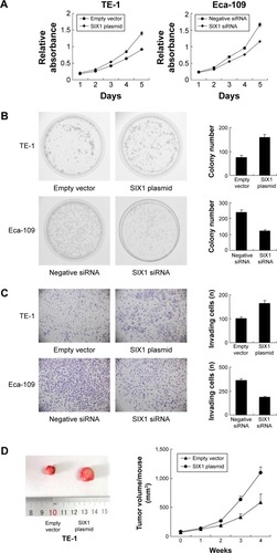
Figure 4 SIX1 regulates radiosensitivity and ERK/AKT signaling.
Notes: (A) AnnexinV/propidium iodide analysis showed, after 4 Gy X-ray treatment, a significant decrease in apoptosis rate in TE-1 cells transfected with SIX1. Transfection with SIX1 siRNA increased the apoptosis rate in Eca-109 cell line. (B) SIX1 transfection upregulated cyclin E, matrix metalloproteinase-2 (MMP-2), Bcl-2, p-extracellular signal-regulated kinase (p-ERK), and p-AKT and downregulated Bim and cleaved caspase-3. SIX1 depletion showed the opposite effects.
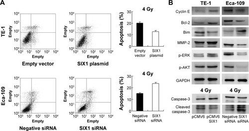
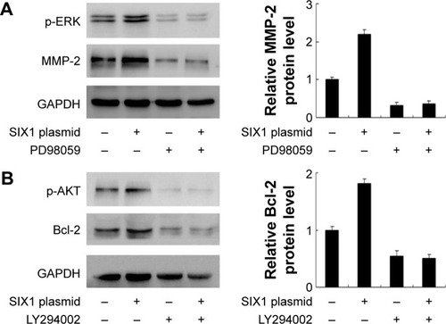
Figure 5 SIX1 regulates matrix metalloproteinase-2 (MMP-2) and Bcl-2 through extracellular signal-regulated kinase (ERK) and AKT signaling.
Notes: (A) ERK inhibitor PD98059 (20 μM) was used to treat TE-1 cell line. Inhibition of ERK signaling reduced p-ERK expression. PD98059 also blocked the role of SIX1 on MMP-2 production. Quantification of protein expression was performed using ImageJ software. (B) LY294002 (10 μM) was used to block AKT signaling in TE-1 cell line. LY294002 treatment could block SIX1 induced change of Bcl-2. Quantification of protein expression was performed using ImageJ software.