
Figures & data
Table 1 Association between the expression of Kindlin proteins and various clinicopathologic features of osteosarcoma patients
Figure 1 Kindlin gene and protein expression levels in human osteosarcoma tissues.
Notes: (A) Kindlin-1 and Kindlin-2 mRNA levels in osteosarcoma tissues were both significantly higher than those in adjacent noncancerous tissues (both P<0.01), whereas Kindlin-3 mRNA levels in osteosarcoma tissues were significantly lower than those in adjacent noncancerous tissues (P<0.05). (B) Kindlin-1 protein levels in osteosarcoma tissues were significantly higher than those in adjacent noncancerous tissues (P<0.01). (C) Kindlin-2 protein levels in osteosarcoma tissues were significantly higher than those in adjacent noncancerous tissues (P<0.01). (D) Kindlin-3 protein levels in osteosarcoma tissues were significantly lower than those in adjacent noncancerous tissues (P<0.01). The mRNA and protein levels of Kindlins were detected by quantitative real-time PCR and Western blot analyses. T and N refer to osteosarcoma and adjacent noncancerous tissues, respectively.
Abbreviations: GAPDH, glyceraldehyde 3-phosphate dehydrogenase; PCR, polymerase chain reaction.
Abbreviations: GAPDH, glyceraldehyde 3-phosphate dehydrogenase; PCR, polymerase chain reaction.
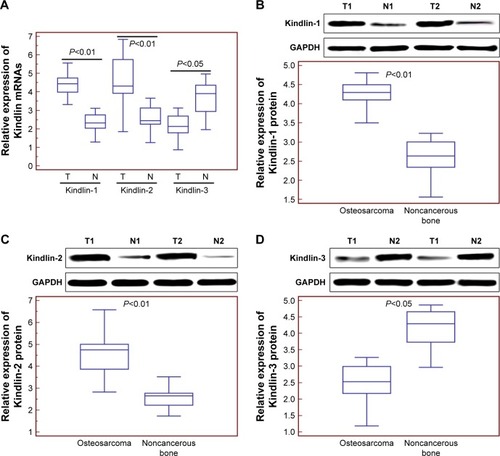
Figure 2 Subcellular localizations and expression patterns of Kindlin proteins in human osteosarcoma tissues.
Notes: The positive Kindlin-1 (A) Kindlin-2 (C) and Kindlin-3 (E) protein immunostainings were localized in the cytoplasm, nucleus and cytoplasm, respectively, of tumor cells in primary osteosarcoma tissues. Statistically, the expression levels of Kindlin-1 (B) and Kindlin-2 (D) proteins in osteosarcoma tissues were both significantly higher, but that of Kindlin-3 protein (F) was dramatically lower than those in noncancerous bone tissues. The subcellular localizations and expression patterns of Kindlins were examined by immunohistochemistry.
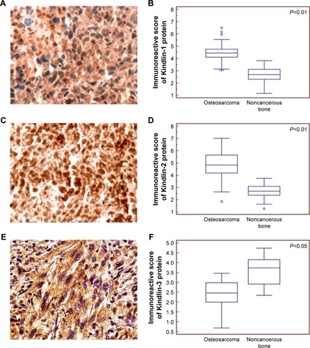
Figure 3 Kaplan–Meier overall (A–C) and disease-free (D–F) survival curves for osteosarcoma patients based on Kindlin-1 (A and D), Kindlin-2 (B and E) and Kindlin-3 (C and F) protein expression.
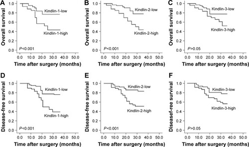
Table 2 Multivariate survival analysis of overall and disease-free survivals in 100 patients with osteosarcomas