Figures & data
Figure 1 Pelvic CT shows the lymph node metastasis. (A) Before therapy (August 17, 2015). (B) Before therapy (October 22, 2015), the lymph node was bigger than 2 months earlier. (C) After 3 months of apatinib treatment (March 7, 2016), the lymph node was smaller than 4 months earlier. (D) After 9 months of apatinib treatment (August 30, 2016), the lymph node was smaller than 5 months earlier. Red arrows indicate the lymph node metastasis.
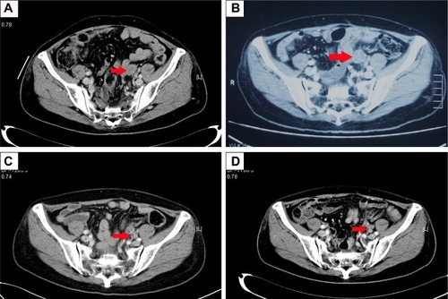
Figure 2 Pelvic CT shows the metastatic mass in front of the rectum. (A) Before therapy (August 17, 2015). (B) Before therapy (October 22, 2015), the mass was bigger than 2 months earlier (C) After 3 months of apatinib treatment (March 7, 2016), the mass was smaller than 4 months earlier. (D) After 9 months of apatinib treatment (August 30, 2016), the mass was smaller than 5 months earlier. Red arrows indicate the metastatic mass in front of the rectum.
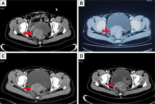
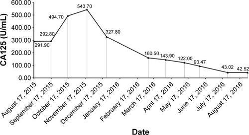
Figure 3 The serum CA125 level during the treatment with apatinib.