Figures & data
Figure 1 The expression of Axl in tissues.
Notes: (A) A representative image of Axl-positive immunohistochemical staining in WT tissues. (B) A representative image of Axl-negative immunohistochemical staining in WT tissues. (C) The Axl expression in WT tissues and adjacent non-tumor tissues by WB. (D) The expression of Axl mRNA in paired WT tissues and adjacent non-tumor tissues by qPCR. Data are shown as mean ± SD. *P<0.05.
Abbreviations: WT, Wilms’ tumor; WB, Western blotting; mRNA, messenger RNA; qPCR, quantitative polymerase chain reaction; SD, standard deviation; T, tumor; N, non-tumor.
Abbreviations: WT, Wilms’ tumor; WB, Western blotting; mRNA, messenger RNA; qPCR, quantitative polymerase chain reaction; SD, standard deviation; T, tumor; N, non-tumor.
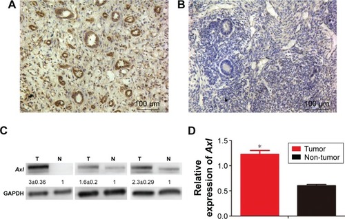
Table 1 Axl expression in WT and matched adjacent noncancerous tissues
Table 2 The relationship between the clinicopathological features and Axl expression of WT patients
Table 3 Univariate Cox analysis of potential prognostic factors in WT patients
Figure 2 The effect of Axl on proliferation of WT cells in vitro.
Notes: (A) The Axl expression of control, sh-Axl and sh-Axl + Gas6 by RT-qPCR. (B) The cell viability assessed by MTT assays in the three groups. (D) The images of clone-forming assays of the three groups. (C) The statistical histogram of clone assays. Data are shown as mean ± SD. *P<0.05, **P<0.01.
Abbreviations: WT, Wilms’ tumor; RT-qPCR, real-time quantitative polymerase chain reaction; MTT, methyl-thiazolyl-tetrazolium; SD, standard deviation.
Abbreviations: WT, Wilms’ tumor; RT-qPCR, real-time quantitative polymerase chain reaction; MTT, methyl-thiazolyl-tetrazolium; SD, standard deviation.
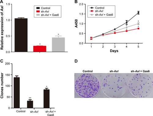
Figure 3 The effect of Axl on migration and invasion of WT cells in vitro.
Notes: (A) The images of wound-healing assays in the three groups. (B) The images of transwell assays in the three groups. (C) The statistical histogram of wound-healing assays. (D) The statistical histogram of transwell assays. Data are shown as mean ± SD. *P<0.05. Scale bar: 100 µm.
Abbreviations: WT, Wilms’ tumor; SD, standard deviation.
Abbreviations: WT, Wilms’ tumor; SD, standard deviation.
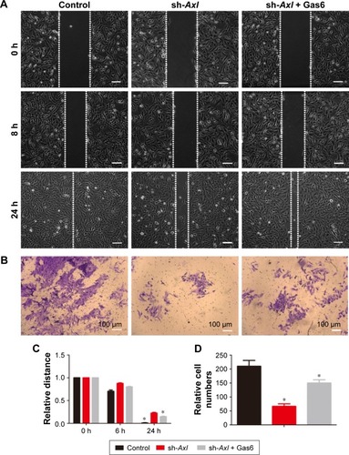
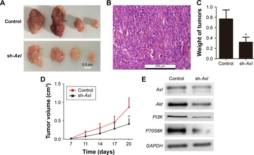
Figure 4 The effect of Axl on tumorigenicity of WT cells in vivo.
Notes: (A) The images of tumors formed in control and sh-Axl WT cells. Scale bar: 0.5 cm. (B) The tumor section with HE staining under microscopy. Scale bar: 200 µm. (C) The statistical histogram of tumor weights. (D) The growth curve of tumors. (E) The expression of proteins Axl, Akt, PI3K and P70S6K by Western blotting. Data are shown as mean ± SD. *P<0.05.
Abbreviations: WT, Wilms’ tumor; HE, hematoxylin and eosin; SD, standard deviation.
Abbreviations: WT, Wilms’ tumor; HE, hematoxylin and eosin; SD, standard deviation.