Figures & data
Figure 1 miR-204-5p is down-regulated in melanoma tissues and cells, and confers a protective effect that improves the prognosis of patients with melanoma. (A) miR-204-5p levels were analyzed in malignant melanoma tissues and benign nevi tissues. (B) The expression of miR-204-5p was analyzed in metastatic melanoma tissues and primary melanoma tissues. (C) The overall survival curves of melanoma patients with high miR-204-5p expression and low miR-204-5p expression. (D) The miR-204-5p expression profile in human melanoma cell lines (A375, WM35, SK-MEL-2 and SK-MEL-5) and human epidermal melanocytes (HEMa-LP). *P<0.05, **P<0.01, ***P<0.001.
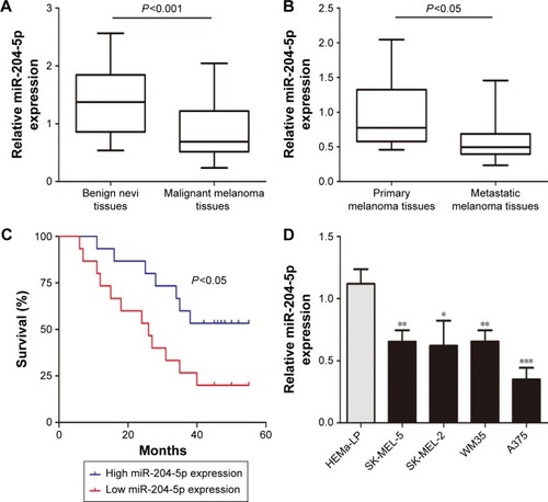
Figure 2 miR-204-5p acts as a tumor suppressor in melanoma. (A) Transfection efficiency of the miR-204-5p mimic was determined by PCR. (B) The proliferative ability of A375 cells was measured by a CCK-8 assay. (C and D) Effect of miR-204-5p on apoptosis of A375 cells was assessed by flow cytometry. (E and F) The effect of miR-204-5p on the invasive capacity of melanoma cells was assessed by a Transwell assay. (G and H) The effect of miR-204-5p on the migratory ability of melanoma cells was assessed by the scratch wound assay. *P<0.05, **P<0.01, ***P<0.001.
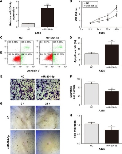
Figure 3 MMP9 and BCL2 are the direct functional targets of miR-204-5p. (A) The binding sites of miR-204-5p within the 3′-UTR of MMP9 and BCL2 were predicted by miRanda. (B) Overexpression of miR-204-5p suppressed luciferase activity in cells with the WT plasmid but did not cause a significant change in A375 cells with the Mut plasmid. (C) Western blots identified MMP9 and BCL2 protein expression changes following transfection with a miR-204-5p mimic or NC; GAPDH was used as a control. (D) PCR to assess the mRNA level of MMP9 and BCL2 in A375 cells following transfection with a miR-204-5p mimic. (E) The heat map shows that MMP9 and BCL2 are negatively correlated with miR-204-5p in thirty malignant melanoma tissues. (F) The Pearson correlation of miR-204-5p and MMP9 and BCL2 expression in thirty malignant melanoma tissues. **P<0.01.
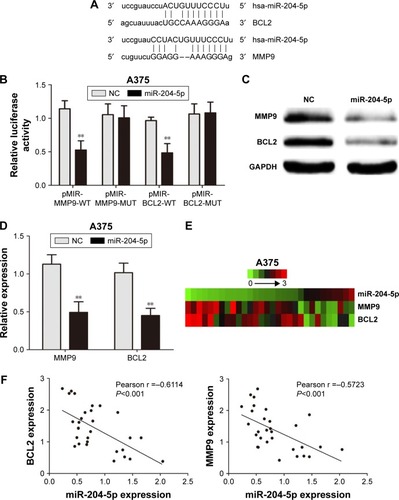
Figure 4 Expression of MMP9 and BCL2 attenuates the effects of miR-204-5p in melanoma. (A) Western blots identified MMP9 and BCL2 protein expression changes following transfection with miR-204-5p alone or in combination with MMP9 and BCL2. (B) BCL2 plasmid reversed the effect of miR-204-5p on the proliferative ability of A375 cells. (C and D) Expression of BCL2 reversed miR-204-5p-induced apoptosis as assessed by flow cytometry. (E and F) The effect of miR-204-5p on the invasive ability of A375 cells was largely abrogated by the MMP9 plasmid. (G and H) The MMP9 plasmid reversed the effect of miR-204-5p on the migratory ability of A375 cells. *P<0.05, **P<0.01.
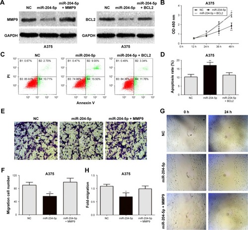
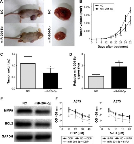
Figure 5 miR-204-5p inhibits melanoma growth in vivo and increases sensitivity of melanoma cells to chemotherapy. (A) Tumor formation in nude mice and the excision tumor of A375 xenografts. (B) Difference in tumor volume between the NC group and the miR-204-5p group. (C) The tumor weight of excision tumor. (D) PCR identified miR-204-5p expression changes. (E) Western blots identified MMP9 and BCL2 protein expression changes. (F) A dose-dependent 5-FU and DDP treatment was conducted on miR-204-5p mimic- or NC-transfected melanoma cells. The proliferative ability of A375 cells was measured by CCK-8 assay. *P<0.05, **P<0.01.