Figures & data
Table 1 Primary antibodies used in Western blot and IHC experiments
Figure 1 Expression of PIK3CA was evaluated using IHC in both DLBCL and RH tissues.
Notes: (A) Weak positive immunostaining of PIK3CA in cells dispersed in and out of the germinal center of RH lymph node. The sublocalization of positive immunostaining of PIK3CA was both cytoplasmic and membranous. (B–D) Negative, weak positive and strong positive immunostaining of PIK3CA in DLBCL tissues (envision method; magnification fold: ×400).
Abbreviations: IHC, immunohistochemistry; DLBCL, diffuse large B cell lymphoma; RH, reactive hyperplasia.
Abbreviations: IHC, immunohistochemistry; DLBCL, diffuse large B cell lymphoma; RH, reactive hyperplasia.
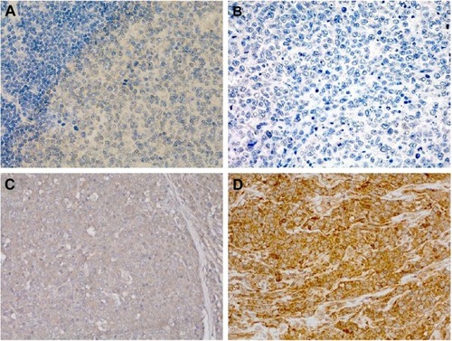
Table 2 Clinicopathological significance of PIK3CD expression in DLBCL
Figure 2 Overall prognostic significance of PIK3CA expression, which was analyzed using the Kaplan–Meier survival curve in DLBCL.
Notes: Of all the patients diagnosed with DLBCL, totaling 205, there were only 101 cases retrieved with the overall prognosis information. Based on the expression status of PIK3CA, 101 cases with prognosis were categorized into two subcohorts, those with low expression of PIK3CA and those with high expression of PIK3CA, with the number of cases being 88 and 23, respectively. The statistical significance was analyzed using the log-rank test.
Abbreviation: DLBCL, diffuse large B cell lymphoma.
Abbreviation: DLBCL, diffuse large B cell lymphoma.
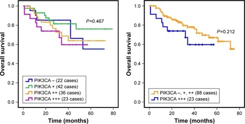
Figure 3 Knockdown of PIK3CA was pronouncedly capable of preventing proliferation and promoting apoptosis, rendering the cell-cycle arrest at the G1 phase in OCI-LY8 cells.
Notes: (A) The basal expression of PIK3CA and p-AKT was detected in six different kinds of DLBCL cell lines that were involved in the study. The molecular weights of PIK3CA, p-AKT and GAPDH were 110, 60 and 36 kDa, as indicated by the manufacturer’s instruction. (B) Based on the successfully stable knockdown of PIK3CA (inserted immunoblotting of PIK3CA), proliferative variation was monitored using the CCK-8 approach. The proliferation of OCI-LY8 cells whose PIK3CA was stably knocked down was remarkably suppressed compared with OCI-LY8 transfected with control vector. (C) Cell apoptosis was assayed using flow cytometry before and after stable knockdown of PIK3CA in OCI-LY8. (D) Quantitative assay of cell late apoptosis. (E) Cell-cycle variation was evaluated using flow cytometry before and after stable knockdown of PIK3CA in OCI-LY8. (F) Quantitative assay of cell-cycle analysis. All the experiments were performed independently in triplicate, and representative figures are shown here. All the data were expressed as mean ± SEM. Two-tailed independent sample t-test was used to analyze the statistical difference. P-value was considered as statistically significant when <0.05 in comparison with the control group.
Abbreviations: p-AKT, phosphorylated AKT; DLBCL, diffuse large B cell lymphoma; SEM, standard error of mean; shRNA, short hairpin RNA interference; 7-AAD, 7-aminoactinomycin D.
Abbreviations: p-AKT, phosphorylated AKT; DLBCL, diffuse large B cell lymphoma; SEM, standard error of mean; shRNA, short hairpin RNA interference; 7-AAD, 7-aminoactinomycin D.
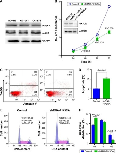
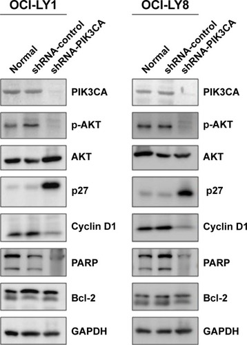
Figure 4 Expression variation of cell cycle- and apoptosis-relevant proteins was detected before and after stable knockdown of PIK3CA in OCI-LY1 and OCI-LY8 cells.
Notes: GAPDH was used as an internal loading control; all the blots were probed and re-probed with primary antibodies on the same membrane. On the basis of successful stable knockdown of PIK3CA, p-AKT, downstream target of PI3K signal pathway, as well as p27, PARP, Bcl-2 and cyclin D1 expression variations were detected using rabbit IgG mono-antibodies. The experiments were done independently in triplicate, and representative figures are presented.
Abbreviations: p-AKT, phosphorylated AKT; shRNA, short hairpin RNA interference.
Abbreviations: p-AKT, phosphorylated AKT; shRNA, short hairpin RNA interference.