
Figures & data
Figure 1 Computed tomography scan of 52-year-old man with mucinous carcinoma in rectum.
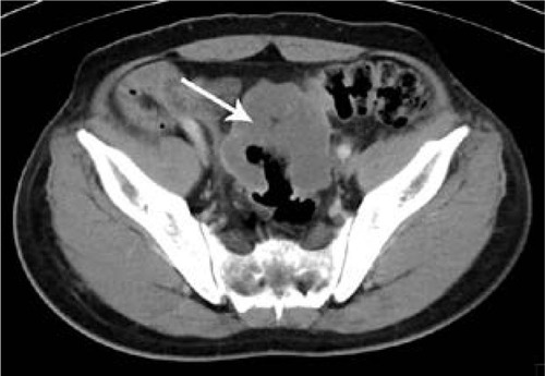
Figure 2 Computed tomography scan of 55-year-old man with nonmucinous carcinoma in rectum.
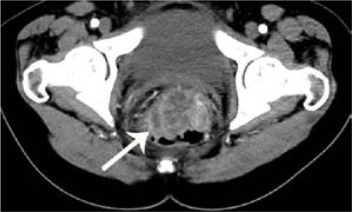
Figure 3 Computed tomography scan of 29-year-old woman with signet-ring cell carcinoma of the rectum.
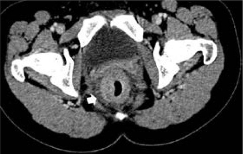
Figure 4 Computed tomography scan of 32-year-old man with signet-ring cell carcinoma of the sigmoideum.
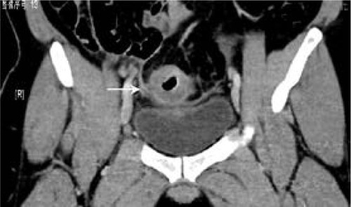
Figure 5 Computed tomography scan of 53-year-old woman with mucinous carcinoma in cecum and proximal ascending colon.
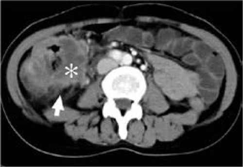
Figure 6 Computed tomography scan of 69-year-old woman with mucinous carcinoma in descending colon.
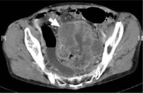
Figure 7 Computed tomography scan of 59-year-old woman with signet-ring cell carcinoma of the rectum.
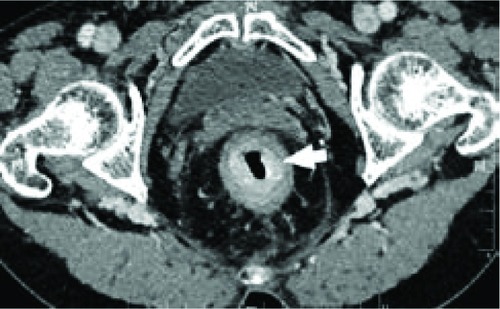
Figure 8 Computed tomography scan of 44-year-old man with mucinous carcinoma in descending colon.
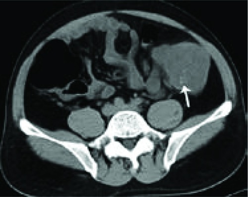
Table 1 Clinical characteristics and computed tomography findings of patients with MAC, SC, and AC