Figures & data
Figure 1 Initial histology and CT findings.
Notes: (A) Microphotograph of adenocarcinoma, acinopapillary subtype. H&E staining, magnification ×40. (B) Axial CT in lung window with solitary nodule in left upper lobe (arrow). (C) Axial CT of abdomen shows solitary liver metastasis (arrow) close to ablation zone in eighth segment. (D) Follow-up CT revealed multiple liver metastases (arrowheads).
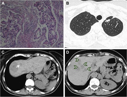
Figure 2 Subsequent CT findings.
Notes: (A) Follow-up CT after 2 months’ treatment shows only two small liver metastases (arrowheads). (B and C) Follow-up CTs of abdomen and thorax from March 2009 show no metastases. (D) Follow-up CT of abdomen from 2012 without liver metastases. (E and F) Follow-up CTs of thorax from 2010 and 2012 show no lung metastases.
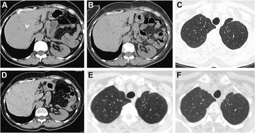
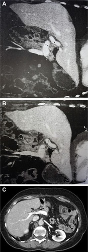
Figure 3 CT finding of disease progression on erlotinib treatment.
Notes: (A) Enlarged lymph nodes of the liver hilum – about 5 cm in diameter (arrows). (B) Follow-up after six cycles of gemcitabine–cisplatin chemotherapy; nearly complete response (arrows). (C) Contrast-enhanced axial CT of abdomen shows small hypodenze node in the liver hilim is unchanged compared with previous CT; nearly complete response (arrows).