Figures & data
Table 1 Clinicopathological characteristics of dogs with primary brain tumors and normal controls
Figure 1 Immunohistochemical evaluation of uPAR expression in control canine brain tissues (A–F) and brain tumors (G–R).
Abbreviations: GBM, glioblastoma; H&E, hematoxylin and eosin; NB, normal brain (cerebral cortex); NCP, normal choroid plexus; NM, normal meninges; uPAR, urokinase plasminogen activator receptor; CII, grade II choroid plexus tumor; OligoIII, grade III oligodendroglioma; MenII, grade II meningioma; MenI, grade I meningioma.
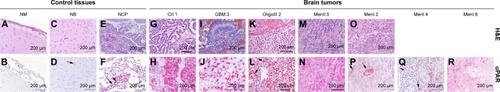
Figure 2 (A) Representative Western blots of uPAR protein in canine normal brain tissues and brain tumors. The numeral after each tumor type and grade indicates the specific sample depicted. (B) uPAR protein expression compared with β-actin concentrations in normal canine brain tissues and tumors.
Abbreviations: GBM, glioblastoma; NM, normal meninges; NB, normal brain (cerebral cortex); NCP, normal choroid plexus; uPAR, urokinase plasminogen activator receptor; MenI, grade I meningioma; MenII, grade II meningioma; CII, grade II choroid plexus tumor; OligoII, grade II oligodendroglioma; OligoIII, grade III oligodendroglioma; AstroII, grade II astrocytoma; AstroIV, grade IV astrocytoma.
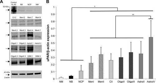
Figure 3 Expression of urokinase plasminogen activator receptor (uPAR) mRNA in canine primary brain tumors as determined by real-time quantitative polymerase chain reaction.
Abbreviations: NB.Cont, normal brain control; MN.Cont, normal meninges control; NCP.Cont, normal choroid plexus control; CII, grade II choroid plexus tumor; CIII, grade III choroid plexus tumor; MenI, grade I meningioma; MenII, grade II meningioma; AstroII, grade II astrocytoma; AstroIII, grade III astrocytoma; GBM, glioblastoma; OligoII, grade II oligodendroglioma; OligoIII, grade III oligodendroglioma.
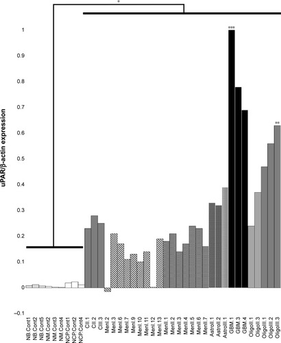
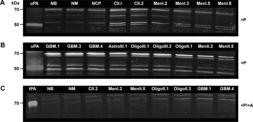
Figure 4 uPA activity in normal canine brain tissues and brain tumors as determined with casein–plasminogen zymography.
Abbreviations: NB, normal brain; NCP, normal choroid plexus; NM, normal meninges; +P, plasminogen-treated gel; MenI, grade I meningioma; MenII, grade II meningioma; CII, grade II choroid plexus tumor; OligoII, grade II oligodendroglioma; OligoIII, grade III oligodendroglioma; AstroII, grade II astrocytoma; AstroIII, grade III astrocytoma; GBM, glioblastoma; +P/+A, plasminogen and amiloride-treated gel; tPA, purified human tissue plasminogen activator control; uPA, urokinase-type plasminogen activator control.