
Figures & data
Figure 1 Abdominal computed tomography findings.
Notes: Computed tomography scan showing a well-demarcated tumor (arrows) in the pancreas head, with heterogeneous enhancement: axial view (A) and intravenous contrast-enhanced axial (B), sagittal (C), and coronal (D) views.
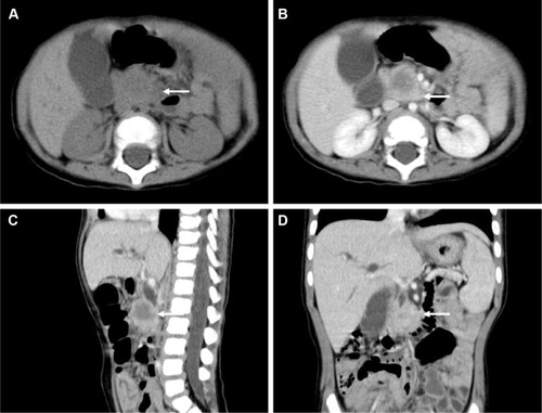
Figure 2 Histopathology of the pancreatic tumor.
Notes: Gross pathology showing a yellowish-gray, firm, well-circumscribed tumor (A). Histological findings showing “patternless pattern” arranged spindle tumor cells and thick collagen (B) (original magnification ×40), and infiltration of the submucosa of the duodenum (C) (original magnification ×40). Immunohistochemical staining demonstrating strong CD34 positivity (D) (original magnification ×100).
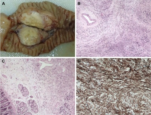
Table 1 Clinicopathological characteristics of pancreatic solitary fibrous tumor in English literature