Figures & data
Table 1 Sequences of siRNAs targeting Foxj2
Figure 1 Expression pattern and prognosis role of Foxj2 in NPC tissues.
Notes: (A) Immunohistochemistry analysis (1:50, ab22857) of Foxj2 expression in NPC tissues. Original magnifications: ×200 (a–c); ×400 (d–f) (B) Protein levels of Foxj2 in NPC (ie, tumor tissue T) and noncancerous nasopharyngeal tissues (labeled “N”) by Western blotting. (C) Quantitative results of Western blotting. β-actin was used as a loading control. The same experiment was repeated at least 3 times. *P<0.05. Data are presented as mean ± SD.
Abbreviation: NPC, nasopharyngeal carcinoma.
Abbreviation: NPC, nasopharyngeal carcinoma.
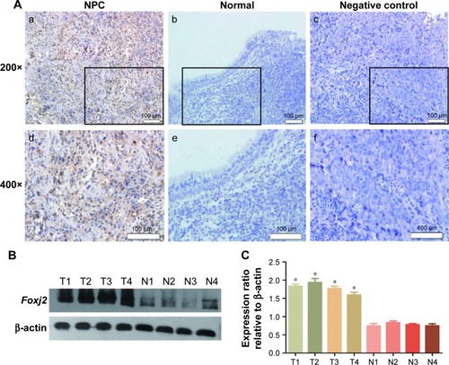
Table 2 The association between Foxj2 expression and clinicopathological features in 57 NPC patients
Figure 2 Prognosis role of Foxj2 in NPC.
Notes: Kaplan–Meier survival curves of NPC patients based on Foxj2 expression status; P<0.05, log-rank test.
Abbreviation: NPC, nasopharyngeal carcinoma.
Abbreviation: NPC, nasopharyngeal carcinoma.
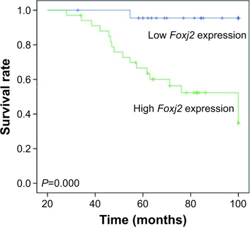
Table 3 Survival status and clinicopathological parameters in 57 human NPC tissues
Table 4 Contribution of various potential prognostic factors to survival, determined by Cox regression analysis, in 57 human NPC tissues
Figure 3 Expression of Foxj2 in NPC cells.
Notes: (A, B) Western blot analysis of Foxj2 expression in 4 kinds of NPC cell lines (CNE-1, CNE-2, 5-8F, and 6-10B) and in NP-69 cell line. (C, D) The expression of Foxj2 was suppressed using siRNAs, and interference efficiency was detected by Western blotting. β-actin was used as control. *P<0.05. Data are presented as mean ± SD.
Abbreviations: NPC, nasopharyngeal carcinoma; siRNA, small interfering RNA.
Abbreviations: NPC, nasopharyngeal carcinoma; siRNA, small interfering RNA.
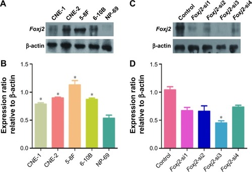
Figure 4 Knockdown of Foxj2 reduces CNE-2 cell proliferation.
Notes: (A) IHC analysis (1:50, ab22857) of Foxj2 expression in T1–T2 and T3–T4 NPC tissues. Original magnifications: ×200 (a–b); ×400 (c–d); (e) percentage expression of Foxj2. (B, C) CCK-8 assay was used to determine cell proliferation of CNE-2 cells treated with Foxj2 siRNA for the indicated time. (D) Cell cycle distribution analyzed by flow cytometry in CNE-2 cells after Foxj2 downregulation. The data are presented as mean value ± SEM (compared with the control). The same experiment was repeated at least 3 times. *P<0.05; ***P<0.001. Data are presented as mean ± SD.
Abbreviations: CCK-8, cell counting kit 8; IHC, immunohistochemistry; NPC, nasopharyngeal carcinoma; OD, optical density; siRNA, small interfering RNA.
Abbreviations: CCK-8, cell counting kit 8; IHC, immunohistochemistry; NPC, nasopharyngeal carcinoma; OD, optical density; siRNA, small interfering RNA.
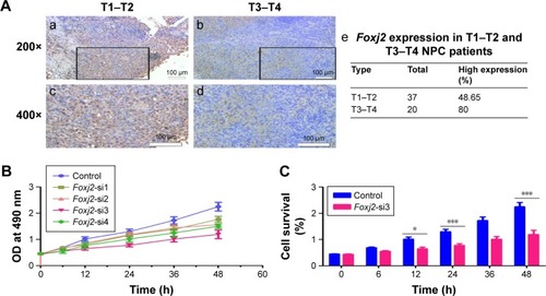
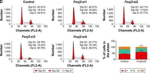
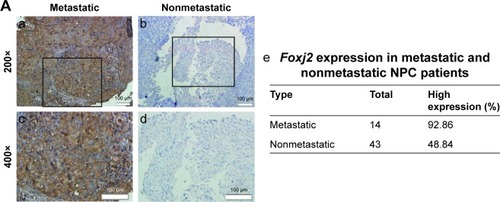
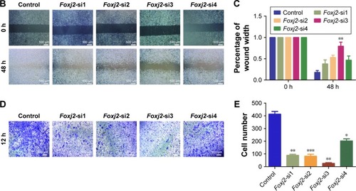
Figure 5 Knockdown of Foxj2 decreases the migration of CNE-2 cells.
Notes: (A) IHC analysis (1:50, ab22857) of Foxj2 expression in metastatic and nonmetastatic NPC tissues. Original magnifications: ×200 (a–b); ×400 (c–d); (e) percentage expression of Foxj2. (B) CNE-2 cells transfected with Foxj2 siRNAs showed a slower migration rate than those transfected with the control siRNA. Migration of cells to the wound was visualized at 0 and 48 h with a microscope (original magnification: ×200). (C) The histogram shows the relative migration distance of (B). (D–E) Knockdown of Foxj2 in CNE-2 cells inhibited cell migration, as determined by transwell assays. Number of cells migrating through the membrane was counted in 10 fields under ×20 objective lens. Error bars, SD. n=3; *P<0.05, **P<0.01, ***P<0.001. The same experiment was repeated at least 3 times.
Abbreviations: IHC, immunohistochemistry; NPC, nasopharyngeal carcinoma; siRNA, small interfering RNA.
Abbreviations: IHC, immunohistochemistry; NPC, nasopharyngeal carcinoma; siRNA, small interfering RNA.