Figures & data
Figure 1 Positron emission tomography–computed tomography examination of metabolically active sternal tumor (white circle: maximum standardized uptake value of fluorine 18 fluorodeoxyglucose =3.5).
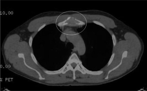
Figure 2 3D model of the implant prepared based on software program including the date of the patient’s computed tomography scan.
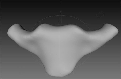
Figure 3 Intraoperative picture: 3D sternal implant filling the defect after tumor resection fixed to bone scaffolds by titanium plates.
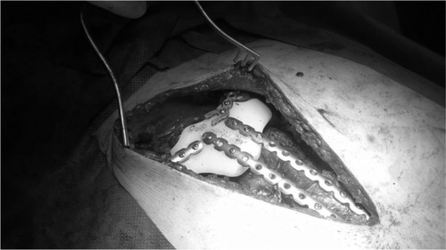
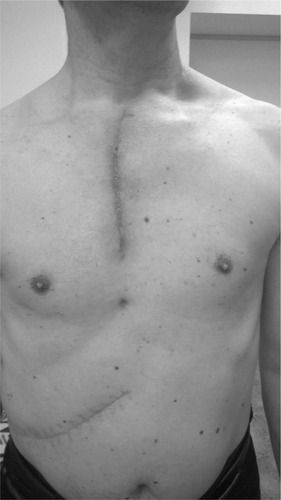
Figure 4 Skin scar 3 months after the operation.