Figures & data
Table 1 The primers used for real-time polymerase chain reaction
Figure 1 Dose-dependent inhibition of cell viability in PANC-1 and PATU-8988 cells, and of epithelial–mesenchymal transition in PATU-8988 cells.
Notes: PANC-1 and PATU-8988 cells were treated with different concentrations of quercetin (0, 20, 40, and 80 μM) for 24 h in the cell viability experiments, and PATU-8988 cells were treated with different concentrations of quercetin (0, 20, 40, and 80 μM) for 24 h in the rest of the experiments. (A, B) Fold changes in cell viability of PANC-1 and PATU-8988 cells. (C) Fold changes in mRNA expression of E-cadherin, N-cadherin, and Vimentin in PATU-8988 cells. PATU-8988 cells were treated with different concentrations of quercetin (0, 20, 40, and 80 μM) for 24 h. (D) The protein expression of E-cadherin, N-cadherin, and Vimentin was measured with a Western blotting assay. β-actin was used as an internal control. (E) Fold changes in mRNA expression of Zeb1, Twist, Slug, and Snail in PATU-8988 cells. (F) The protein expression of Zeb1, Twist, Slug, and Snail was measured with a Western blotting assay in PATU-8988 cells. β-actin was used as an internal control. (G) The immunofluorescence of N-cadherin, Vimentin (green), and the cell nuclei (blue). The data are presented as the mean ± standard error of the mean (SEM); n=3; *P<0.05 versus the 0 μM group.
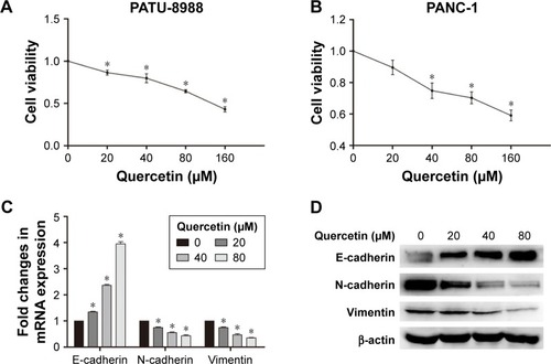
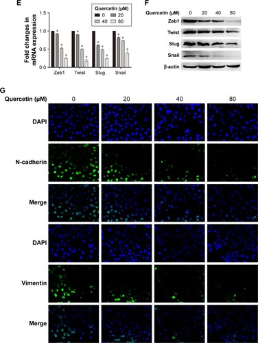
Figure 2 Quercetin inhibits invasion and metastasis of PATU-8988 cells in a dose-dependent manner.
Notes: PATU-8988 cells were treated with different concentrations of quercetin (0, 20, 40, and 80 μM) for 24 h. (A) Fold changes in mRNA expression of MMP-2 and MMP-7 in PATU-8988 cells. (B) The protein expression of MMP-2 and MMP-7 was measured with a Western blotting assay. β-actin was used as an internal control. (C) Quercetin inhibited the invasion and migration capacity of PATU-8988 cells. (D) Image of the wound healing assay and the average width of the wound area. The data are presented as the mean ± standard error of the mean (SEM); n=3; *P<0.05 versus the 0 μM group.
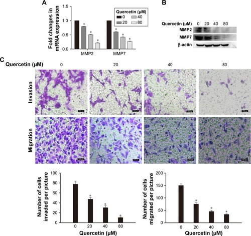
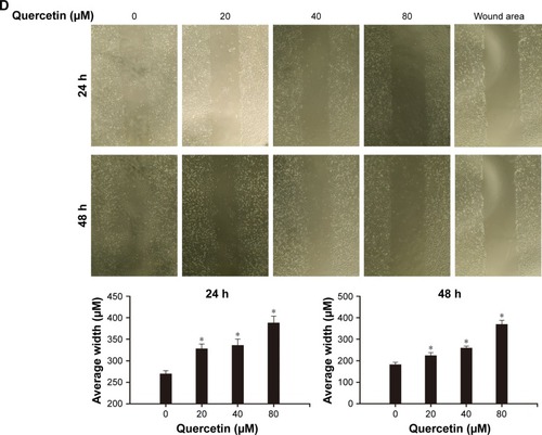
Figure 3 Quercetin inhibits the STAT3 signaling pathway and IL-6-induced STAT3 signaling.
Notes: (A) The protein expression of STAT3 and p-STAT3 was measured with a Western blotting assay. β-actin was used as an internal control. PATU-8988 cells were treated with different concentrations of quercetin (0, 20, 40, and 80 μM) for 24 h. (B) Morphological changes after IL-6 (100 ng/mL) treatment in PATU-8988 cells. (C) The protein expression of IL-6-induced STAT3 and p-STAT3 was measured with a Western blotting assay. β-actin was used as an internal control. PATU-8988 cells were treated with human recombinant IL-6 (100 ng/mL) for 72 h, and in the last 24 h, quercetin (80 μM) was added. (D) The protein expression of IL-6-induced p-STAT3 in cells treated with quercetin and AG490. β-actin was used as an internal control. Before being treated with quercetin for 24 h, PANC-1 cells were pretreated with AG490 for 30 min.
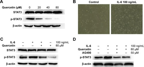
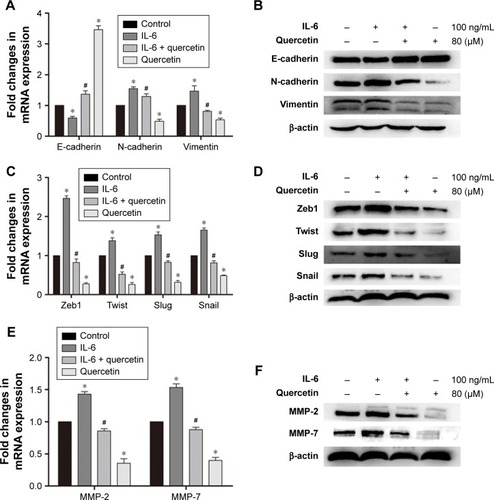
Figure 4 Quercetin inhibits IL-6-induced epithelial–mesenchymal transition and MMP secretion in PATU-8988 cells.
Notes: PATU-8988 cells were treated with human recombinant IL-6 (100 ng/mL) for 72 h, and in the last 24 h, quercetin (80 μM) was added. (A) Fold changes in mRNA expression of E-cadherin, N-cadherin, and Vimentin in PATU-8988 cells. (B) The protein expression of E-cadherin, N-cadherin, and Vimentin was measured with a Western blotting assay. β-actin was used as an internal control. (C) Fold changes in the mRNA expression of Zeb1, Twist, Slug, and Snail in PATU-8988 cells. (D) The protein expression of Zeb1, Twist, Slug, and Snail was measured with a Western blotting assay in PATU-8988 cells. β-actin was used as an internal control. (E) Fold changes in the mRNA expression of MMP-2 and MMP-7 in PATU-8988 cells. (F) The protein expression of MMP-2 and MMP-7 was measured with a Western blotting assay. β-actin was used as an internal control. The data are presented as the mean ± standard error of the mean (SEM); n=3; *P<0.05 versus the 0 μM group; #P<0.05 versus the 20 μM group.