Figures & data
Figure 1 Relative expression of PANDAR in CCA and its clinical significance.
Notes: (A) Relative expression of PANDAR in CCA tissues and adjacent nontumor tissues were determined by qRT-PCR; (B) Relative expression of PANDAR in HIBEC and CCA cell lines were determined by qRT-PCR; (C) Kaplan–Meier survival curves showed that overexpression of PANDAR decreased overall survival in patients with CCA. *P<0.05, **P<0.01, ***P<0.001.
Abbreviations: CCA, cholangiocarcinoma; qRT-PCR, quantitative real-time PCR.
Abbreviations: CCA, cholangiocarcinoma; qRT-PCR, quantitative real-time PCR.
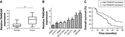
Table 1 Association between PANDAR expression and clinicopathological characteristics of CCA
Table 2 Univariate and multivariate analysis of prognostic factors for overall survival in patients with CCA
Figure 2 Transfection and knockdown efficiency of PANDAR-specific siRNAs and PANDAR depletion retarded cell proliferation and colony formation in CCA cells.
Notes: (A) Transfection efficiency detected by flow cytometry in QBC939 and RBE cells; (B) Relative expression of PANDAR in QBC939 and RBE cells was significantly decreased by three siRNAs specifically targeting PANDAR compared with si-NC; (C) CCK-8 assay showed that silenced PANDAR inhibited cell proliferation of QBC939 and RBE cells; (D) Clonogenic assay showed that silenced PANDAR dramatically impaired the colony formation ability of QBC939 and RBE cells. *P<0.05, **P<0.01.
Abbreviation: CCA, cholangiocarcinoma.
Abbreviation: CCA, cholangiocarcinoma.

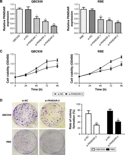
Figure 3 PANDAR depletion increased apoptosis in CCA cells.
Notes: (A) Flow cytometry for apoptosis analysis showed that silenced PANDAR significantly promoted apoptosis of QBC939 and RBE cells compared to negative control siRNA; (B) AO/EB double fluorescence staining assay showed that silenced PANDAR significantly promoted apoptosis of QBC939 and RBE cells compared to negative control siRNA (×200); (C) Relative expression of caspase-3 and caspase-9 in QBC939 and RBE cells were activated by si-PANDAR-2; (D) Western blot analysis showed that knockdown of PANDAR restrained the expression of Bcl-2 and increased Bax expression in QBC939 and RBE cells. *P<0.05, **P<0.01.
Abbreviations: CCA, cholangiocarcinoma; AO/EB, acridine orange/ethidium bromide.
Abbreviations: CCA, cholangiocarcinoma; AO/EB, acridine orange/ethidium bromide.
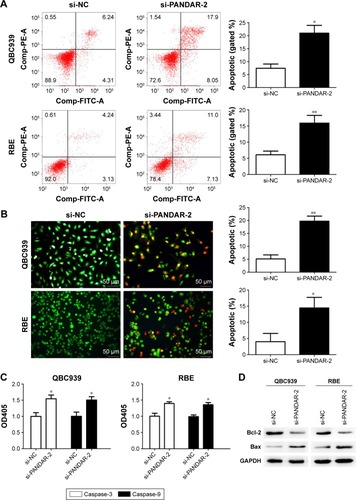
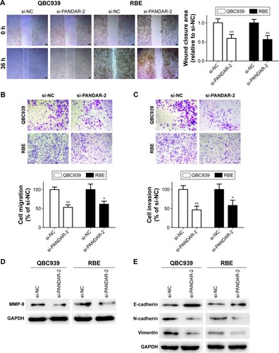
Figure 4 Knockdown of PANDAR impaired migration and invasion capacity in vitro and reversed EMT of CCA cells.
Notes: (A) Wound healing assay showed that silenced PANDAR inhibited migration ability of QBC939 and RBE cells (×50); (B) Transwell migration assay showed that silenced PANDAR inhibited migration capacity of QBC939 and RBE cells (×100); (C) Transwell invasion assay showed that silenced PANDAR inhibited invasion potential of QBC939 and RBE cells (×100); (D) Western blot analysis showed that knockdown of PANDAR restrained the expression of MMP-9 in QBC939 and RBE cells; (E) Knockdown of PANDAR reversed EMT in QBC939 and RBE cells. *P<0.05, **P<0.01.
Abbreviations: CCA, cholangiocarcinoma; EMT, epithelial-to-mesenchymal transition.
Abbreviations: CCA, cholangiocarcinoma; EMT, epithelial-to-mesenchymal transition.