Figures & data
Figure 1 Osimertinib induces apoptosis in CRC cells.
Notes: (A) HCT15 and SW48 cells were treated with increasing concentrations of osimertinib for 72 hours. Cell proliferation was determined by MTS assay. Results were expressed as means ± SD of three independent experiments. (B) HCT15 cells were treated with 10 μg/mL osimertinib with or without z-VAD for 24 hours. Apoptosis was analyzed by Annexin V/PI staining followed by flow cytometry. (C) HCT15 cells were treated with 10 μg/mL osimertinib for 24 hours. Caspase 3/7 activity was determined by fluorogenic analysis. (D) HCT15 cells were treated with 10 μg/mL osimertinib for 24 hours. Cleaved caspase 3 and 9 were analyzed by Western blotting. **P<0.01; ***P<0.001 compared with the vehicle control group.
Abbreviations: CRC, colorectal cancer; MTS, 3-(4,5-dimethylthiazol-2-yl)-5-(3-carboxymethoxyphenyl)-2-(4-sulfophenyl)-2H-tetrazolium; PI, propidium iodide.
Abbreviations: CRC, colorectal cancer; MTS, 3-(4,5-dimethylthiazol-2-yl)-5-(3-carboxymethoxyphenyl)-2-(4-sulfophenyl)-2H-tetrazolium; PI, propidium iodide.
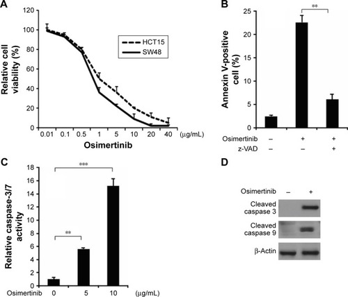
Figure 2 Osimertinib induces p53-independent PUMA induction in CRC cells.
Notes: (A) Parental and p53-KD HCT15 cells were treated with osimertinib at indicated time point. p53 knockdown was confirmed by Western blotting (left). PUMA mRNA induction by osimertinib was analyzed by RT-PCR, with β-actin as a control (right). (B) HCT15 cells were treated with 10 μg/mL osimertinib at indicated time point. Total RNA was extracted, and PUMA mRNA expression was analyzed by semiquantitive RT-PCR. β-Actin was used as a control. (C) HCT15 cells treated with 10 μg/mL osimertinib at indicated time point. PUMA protein levels were analyzed by Western blotting. (D) SW48 cells treated with 10 μg/mL osimertinib at indicated time point. PUMA protein levels were analyzed by Western blotting. (E) HCT15 cells treated with 10 μg/mL osimertinib at indicated time point. Indicated protein levels were analyzed by Western blotting.
Abbreviations: CRC, colorectal cancer; p53-KD, p53-Knockdown; PUMA, p53 upregulated modulator of apoptosis; RT-PCR, reverse transcription polymerase chain reaction.
Abbreviations: CRC, colorectal cancer; p53-KD, p53-Knockdown; PUMA, p53 upregulated modulator of apoptosis; RT-PCR, reverse transcription polymerase chain reaction.
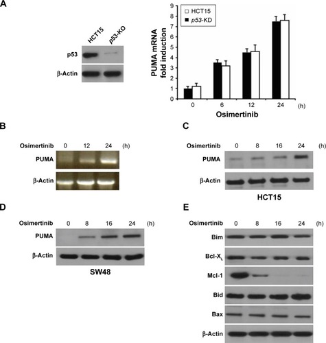
Figure 3 PUMA mediates the anticancer effects of osimertinib through the mitochondrial apoptotic signaling.
Notes: (A) Parental, p53-KD and PUMA-KD HCT15 cells were treated with osimertinib at indicated concentration for 24 hours. PUMA knockdown was confirmed by Western blotting (left). Apoptosis was analyzed by a nuclear fragmentation assay (right). (B) Parental and PUMA-KD cells treated with osimertinib at indicated concentration for 24 hours. Apoptosis was analyzed by Annexin V/PI staining followed by flow cytometry. (C) Parental and PUMA-KD HCT15 cells were treated with osimertinib, and caspase 3/7 activity was determined by fluorogenic analysis. (D) Parental and PUMA-KD cells were treated with 10 μg/mL osimertinib for 24 hours. Cleaved caspase 3 and 9 were analyzed by Western blotting. (E) The cytoplasm and mitochondria were fractionated from parental and PUMA-KD HCT15 cells treated with 10 μg/mL osimertinib for 24 hours. The distribution of cytochrome C was analyzed by Western blotting. β-Actin and cytochrome oxidase subunit IV (Cox IV) were analyzed as the control for loading and fractionation. Results in (A), (B) and (C) were expressed as means ± SD of 3 independent experiments. **P<0.01.
Abbreviations: COX, cytochrome oxidase; p53-KD, p53-Knockdown; PUMA, p53 upregulated modulator of apoptosis; PUMA-KD, p53 upregulated modulator of apoptosis-Knockdown; PI, propidium iodide.
Abbreviations: COX, cytochrome oxidase; p53-KD, p53-Knockdown; PUMA, p53 upregulated modulator of apoptosis; PUMA-KD, p53 upregulated modulator of apoptosis-Knockdown; PI, propidium iodide.
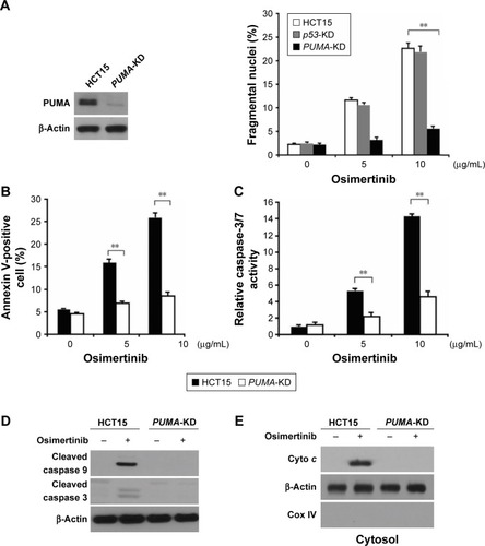
Figure 4 p73 mediates osimertinib-induced PUMA induction.
Notes: (A) HCT15 cells were treated with 10 μg/mL osimertinib at indicated time point. p73, p-p65, E2F1, p-FoxO3a and FoxO3a expression was analyzed by Western blotting. (B) HCT15 cells were treated with 10 μg/mL osimertinib at indicated time point. p73 expression was analyzed by Western blotting. (C) HCT15 cells were transfected with either a control-scrambled siRNA or a p73 siRNA for 24 hours, and then treated with 10 μg/mL osimertinib for 24 hours. p73 and PUMA expression was analyzed by Western blotting. (D) ChIP was performed using anti-p73 antibody on HCT15 cells following osimertinib treatment for 8 hours. ChIP with the control IgG was used as a control. PCR was carried out using primers surrounding the p73 binding sites in the PUMA promoter.
Abbreviations: ChIP, chromatin immunoprecipitation; PCR, polymerase chain reaction; PUMA, p53 upregulated modulator of apoptosis.
Abbreviations: ChIP, chromatin immunoprecipitation; PCR, polymerase chain reaction; PUMA, p53 upregulated modulator of apoptosis.
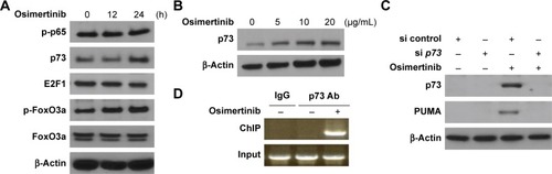
Figure 5 PUMA induction by osimertinib is mediated through AKT inactivation.
Notes: (A) HCT15 cells were treated with osimertinib for 24 hours. Total AKT and phospho-AKT were analyzed by Western blotting. (B) HCT15 cells were trans-fected with Active-AKT plasmid for 6 hours, and then treated with 10 μg/mL osimertinib for 24 hours. PUMA, p73 and HA (AKT) were analyzed by Western blotting.
Abbreviation: PUMA, p53 upregulated modulator of apoptosis.
Abbreviation: PUMA, p53 upregulated modulator of apoptosis.
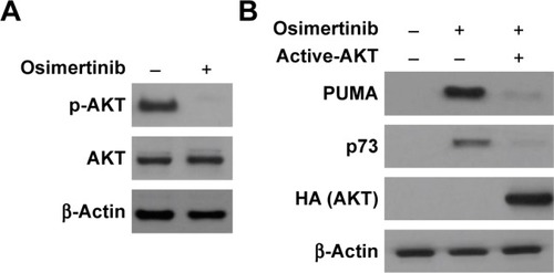
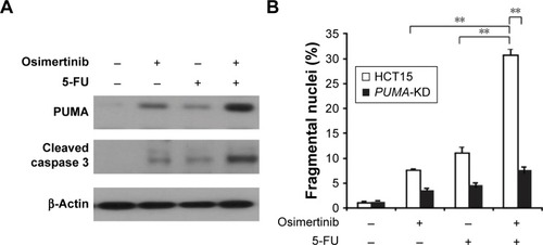
Figure 6 Osimertinib synergizes with 5-FU to induce cell apoptosis via PUMA.
Notes: (A) HCT15 cells were treated with 5 μg/mL osimertinib, 20 mg/L 5-FU, or the combination for 24 hours. PUMA and cleaved-caspase 3 were analyzed by Western blotting. (B) Parental and PUMA-KD HCT15 cells were treated with 5 μg/mL osimertinib, 20 mg/L 5-FU, or the combination for 24 hours. Apoptosis was analyzed by a nuclear fragmentation assay. Results in (B) were expressed as means ± SD of 3 independent experiments. **P<0.01.
Abbreviations: 5-FU, 5-fluorouracil; PUMA, p53 upregulated modulator of apoptosis; PUMA-KD, p53 upregulated modulator of apoptosis-Knockdown.
Abbreviations: 5-FU, 5-fluorouracil; PUMA, p53 upregulated modulator of apoptosis; PUMA-KD, p53 upregulated modulator of apoptosis-Knockdown.