
Figures & data
Figure 1 Hematoxylin–eosin staining and immunohistochemistry in adenocarcinoma before crizotinib treatment.
Notes: (A) Hematoxylin–eosin staining revealed that tumor cells were lung adenocarcinoma (×200). (B) Immunohistochemical examination revealed that tumor cells were positive for monoclonal anti-TTF-1 antibody (×200). (C) Immunohistochemical examination revealed that tumor cells were positive for monoclonal anti-Napsin A antibody (×200).
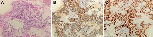
Figure 2 Amplification plot of EML4-ALK-positive by amplification refractory mutation system method.
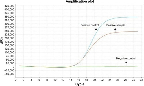
Figure 3 Hematoxylin–eosin staining and immunohistochemistry in small-cell cancer after crizotinib treatment.
Notes: (A) Hematoxylin–eosin staining revealed that tumor cells were lung small-cell cancer (×100). (B) Immunohistochemical examination revealed that tumor cells were positive for monoclonal anti-CD56 antibody (×100). (C) Immunohistochemical examination revealed that tumor cells were positive for monoclonal anti-Syn antibody (×200). (D) Immunohistochemical examination revealed that the tumor cell proliferation index was 98% for monoclonal anti-Ki-67 antibody (×100). (E) Immunohistochemical examination revealed that tumor cells were negative for monoclonal anti-CgA antibody (×100). (F) Immunohistochemical examination revealed that tumor cells were negative for monoclonal anti-PD-L1 antibody (×100).
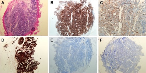
Table 1 Primary antibodies used for immunhistochemical staining
Table 2 Details of the treatment process
Table 3 List of reported small-cell transformations resistant to crizotinib/alectinib/ceritinib/lorlatinib