
Figures & data
Figure 1 The theranostic principle in nuclear medicine involves combining diagnostic imaging and therapy with the same molecule, which is radiolabeled differently, or administered in other dosages. In case of radioiodine therapy (RAI), the radioisotope (131I or 123I) can be directly mediated by the sodium-iodide symporter in the thyroid cells. In other cases, it can be more complex. The image shows a simplified model of a radiopharmaceutical, which consists of a binding molecule that binds the target, and a linking molecule, which binds the radioisotope. Examples of such theranostic molecules are DOTA-TOC, DOTA-TATE, and PSMA-617.
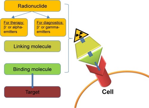
Table 1 Overview of theranostic agents
Figure 2 (A) Initial 131I-planar images of a 74-year-old female with metastatic thyroid cancer (lung, bone, and intracranial soft-tissue metastases, marked with arrows). The tumor marker thyroglobulin (Tg) before radioiodine therapy (RAI) was 572 ng/mL. (B) After two administrations of RAI (cumulative activity: 14.3 GBq), the patient was in complete remission with the Tg at 0.2 ng/mL. The planar images show only a physiological uptake of the radiotracer in the gastrointestinal tract and pharyngeal mucosa (marked with an asterisk).
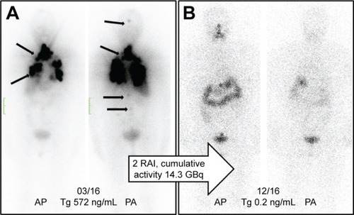
Figure 3 (A) [68Ga]Ga-DOTA-TOC-image* of a male patient with a neuroendocrine tumor (unknown origin) 23 months after the baseline PRRT (three cycles, cumulative activity: 19.6 GBq). The patient had a recurrence of the disease with multiple metastases in the bone, lung, liver, and lymph nodes (marked with arrows). (B) After another course of PRRT (three cycles, cumulative: 43.4 GBq), the PET images showed a partial response.
Abbreviations: PET, positron emission tomography; PRRT, peptide receptor radionuclide therapy.
![Figure 3 (A) [68Ga]Ga-DOTA-TOC-image* of a male patient with a neuroendocrine tumor (unknown origin) 23 months after the baseline PRRT (three cycles, cumulative activity: 19.6 GBq). The patient had a recurrence of the disease with multiple metastases in the bone, lung, liver, and lymph nodes (marked with arrows). (B) After another course of PRRT (three cycles, cumulative: 43.4 GBq), the PET images showed a partial response.](/cms/asset/4473e871-533c-40d5-877e-f63f0d61123b/dott_a_12195610_f0003_c.jpg)
Figure 4 (A) Pre-therapeutic [68Ga]Ga-PSMA-11 images* of a patient with multiple lymph node, peritoneal, and bone metastases (arrows), and history of chemotherapy (first and second line) with enzalutamide and abiraterone. (B) After three cycles of [177Lu]Lu-PSMA-617, the follow-up images showed a very good response with a substantial PSA decline.
Abbreviations: PET, positron emission tomography; PSA, prostate-specific antigen.
![Figure 4 (A) Pre-therapeutic [68Ga]Ga-PSMA-11 images* of a patient with multiple lymph node, peritoneal, and bone metastases (arrows), and history of chemotherapy (first and second line) with enzalutamide and abiraterone. (B) After three cycles of [177Lu]Lu-PSMA-617, the follow-up images showed a very good response with a substantial PSA decline.](/cms/asset/8fea53dd-459d-4e09-b8ed-efd71061cd99/dott_a_12195610_f0004_c.jpg)