Figures & data
Table 1 Breast cancer subtype classifications, based on site of occurrence and/or biomarker status
Table 2 Main molecular alterations in pathways associated with breast cancer and respective targeted therapies
Figure 1 ER genomic pathway.
Notes: (A) E2–ER activates gene expression through direct binding to specific ERE sequences, recruiting coactivators (CoAs) and histone acetyltransferases (HATs) (1); ER and E2 modulate gene expression through interactions with other transcription factors (TFs) (2); ER ligand-independent activation through receptor tyrosine kinase (RTK) signaling (3). (B) In breast cancer, the microenvironment containing fibroblasts and inflammatory and endothelial cells has a critical role in the initiation and progression of tumors, providing growth factors, cytokines, and chemokines that activate ER and upregulate its target genes. (C) ER nongenomic pathway. ER localizes at the cell membrane in caveolae, binds Cav1 and interacts with adaptor proteins/cSrc. This complex activates MAPK and PI3K–Akt pathways (1). E2/GPER at the cell membrane and endoplasmic reticulum activates cSrc signaling, releasing HB-EGF, which binds to EGFR and activates MAPK/PI3K–Akt pathways (2). ER associates with RTKs in response to E2 binding. E2–ER recruits additional coactivator molecules, leading to the activation of RTKs and the downstream kinase pathway (3).
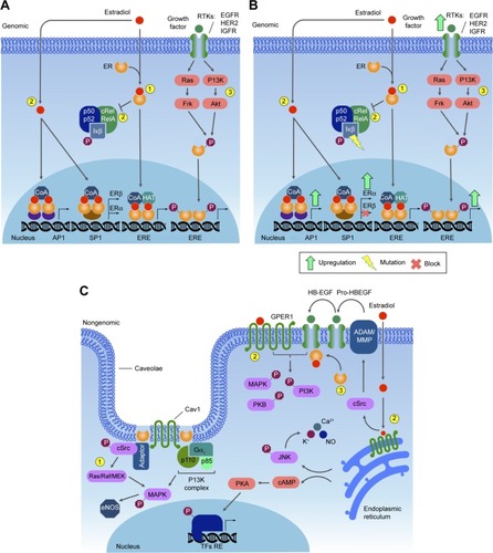
Figure 2 The PI3K signaling pathway.
Notes: (A) Activation of receptor tyrosine kinase (RTK) by a ligand results in phosphorylation of the receptor and adaptor proteins. The PI3K catalytic subunit phosphorylates PIP2 (PtdIns[4,5]–P2), generating PIP3 (PtdIns[3–5]–P3), which recruits Akt and PDKs to the plasma membrane. Akt can be phosphorylated and activated by both PIP3 and PDK1. Activated Akt modulates several transcription factors. (B) In breast cancer, mutations in several PI3K pathway members impact downstream functions, including gene expression.
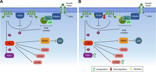
Figure 3 MAPK signaling pathway.
Notes: (A) Specific ligands bind to the transmembrane receptor tyrosine kinase (RTK). The receptor, activated by transphosphorylation, recruits GRB2 and SOS, changing Ras-GDP to active Ras-GTP. Ras-GTP recruits and phosphorylates MAPKKK, which in turn phosphorylates MAPKK and finally MAPK. MAPK is translocated to the nucleus, which will further phosphorylate AP1 transcription factors that will mediate expression of target genes containing a TPA DNA-response element (TRE), like cFos and cJun. (B) In breast cancer, excess extracellular growth factors or mutations in Ras and/or MAPK affect the expression of target genes.
Abbreviation: TPA, tetradecanoyl phorbol acetate.
Abbreviation: TPA, tetradecanoyl phorbol acetate.
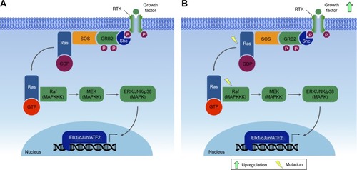
Figure 4 JAK–STAT signaling pathway.
Notes: (A) Specific ligands bind to membrane receptors, leading to dimerization, which promotes JAK activation and transphosphorylation, which in turn phosphorylates STAT, prompting its dimerization. STAT dimers enter the nucleus and binds to specific DNA sequences, activating gene expression. (B) In breast cancer, constitutive activation of certain members of the pathway leads to the overexpression of genes that promote growth and apoptosis resistance.
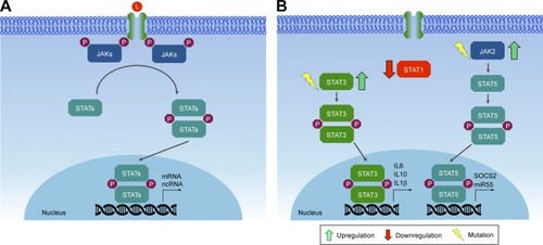
Figure 5 Wnt pathways.
Notes: (A) Planar cell polarity (PCP) Wnt pathway in “off” state, with no ligand bound to Fzd receptor leads to β-catenin proteasome degradation. (B) In the noncanonical β-catenin-independent PCP Wnt pathway, Wnt binds to the Fzd receptor and membrane coreceptors ROR and Ryk. These receptors activate the cytoplasmic signaling protein Dvl, which interacts with Rac and DAAM. Rac activates JNK, while DAAM activates Rho, which activates Rock to regulate cellular cytoskeletal arrangements and actin polymerization. (C) Wnt signaling can also go through the alternative Ca2+-dependent β-catenin-independent pathway, where signaling is mediated by Dvl, G proteins, PKC, CamKII and Cn (calcineurin).
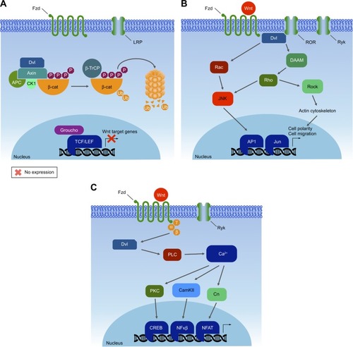
Figure 6 Canonical Wnt signaling pathway.
Notes: (A) Wnt binds to the Fzd receptor and induces the binding of Axin to LRP. The proteasome decouples, allowing β-catenin to stabilize. Accumulated β-catenin translocates to the nucleus, where it binds TCF sites to promote the expression of target genes. (B) A new model of Wnt signaling in which Wnt binds to Fzd receptor and recruits a large protein complex composed of Dvl, Axin, and β-catenin, among others. Subsequently, β-catenin enters the nucleus to regulate gene expression. In breast cancer, both Dvl and β-catenin can be overexpressed due to different mutations, leading to upregulation of a series of target genes.
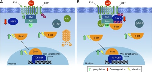
Figure 7 TGFβ signaling in breast cancer progression
Notes: TGFβ modulates cell proliferation through autocrine and paracrine signaling (1). TGFβ binds its receptor on the target cell, signaling through intracellular SMAD to activate transcription of target genes, notably those that effect epithelial–mesenchymal transition (EMT) (2). Snail/Twist family of transcription factors promote transition to the mesenchymal state, corresponding to a shift from epithelial markers, such as E-cadherin and claudin, to mesenchymal markers, such as N-cadherin and vimentin. MiR200f noncoding RNAs antagonize expression of Snail/Twist to promote the epithelial state (3). Migrating mesenchymal cells acquire invasive properties, such as intravasation, vascular survival, and extravasation, and effect a mesenchymal–epithelial transition to contribute to metastatic tumors (4). In breast cancer, TGFβ signaling is active in a subpopulation of CD44+/CD24−/claudin-low breast tumor-initiation cells, which share properties with normal mammary stem cells. These cells express mesenchymal markers, and are enriched after chemotherapy, promoting recurrence of metastatic tumors (5).
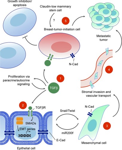
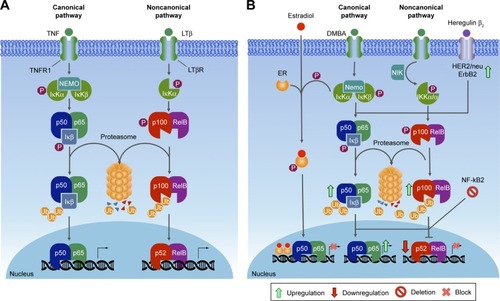
Figure 8 NFκB pathway.
Notes: (A) The canonical pathway starts with binding of TNF to its receptor, activating the IKK complex, which phosphorylates IκBα, promoting its polyubiquitination and further proteasome-dependent degradation. Free NFκB rapidly enters the nucleus, where it can activate the expression of specific genes, and noncanonical pathway induction leads to the activation of NIK, which phosphorylates and activates IKKα, phosphorylating p100, resulting in its ubiquitination and partial processing to p52. (B) In breast cancer ER+ tumors, IκBα phosphorylation is inhibited by estrogen in the canonical pathway. In contrast, some carcinogens like 7,12-dimethylbenz[a]anthracene (DMBA) increase NFκB. In the noncanonical pathway, the deletion of the NFκB2 gene leads to inhibition of target gene expression, while at the same time it promotes an increase in canonical pathway activity.