Figures & data
Figure 1 Gene expression analysis of β-adrenergic receptor expression in DAOY medulloblastoma cells. Total RNA was extracted from medulloblastoma-derived DAOY cells and semi-quantitative RT-PCR performed as described in the Methods section. cDNA amplicons were resolved on an agarose gel in order to confirm a single amplification product.
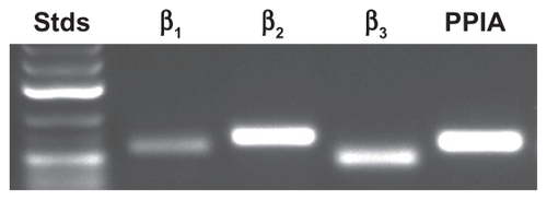
Figure 2 Propranolol inhibits PMA-induced matrix metalloproteinase-9 (MMP-9) secretion in DAOY medulloblastoma cells. Medulloblastoma-derived DAOY cells were serum-starved in the presence of various concentrations of propranolol in combination with vehicle or 1 μM PMA for 18 hours (A), or in the presence of various concentrations of PMA in combination with vehicle or 30 μM propranolol for 18 hours (B). Scanning densitometry was used to quantify the extent of proMMP-9 gelatinolytic activity for each set of data (C, D). Data shown is representative of two independent experiments.
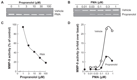
Figure 3 Propranolol reverses PMA-mediated IκB degradation. Medulloblastoma-derived DAOY cells were serum-starved in the presence of various concentrations of propranolol in combination with vehicle or1 μM PMA for 18 hours. A) Lysates were isolated, electrophoresed via sodium dodecylsulfate–polyacrylamide gel electrophoresis and immunodetection of IκB and GAPDH proteins was performed as described in the Methods section. B) Quantification was performed by scanning densitometry of the autoradiograms. Data were expressed as the percent (%) expression of untreated basal conditions.
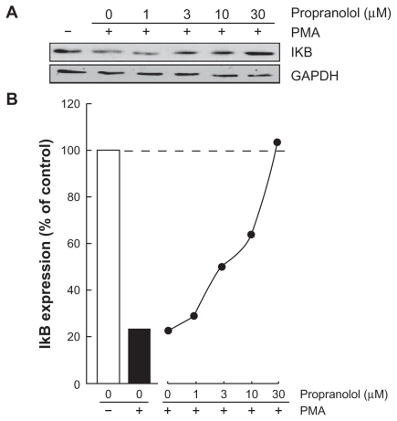
Figure 4 Propranolol inhibits PMA-induced IκB phosphorylation that leads to IκB degradation. A) Medulloblastoma-derived DAOY cells were serum-starved for 30 minutes in the presence of vehicle or 30 μM propranolol. Cells were then incubated for the indicated time with vehicle or 1 μM PMA. Lysates were isolated, electrophoresed via sodium dodecylsulfate–polyacrylamide gel electrophoresis and immunodetection of phosphorylated IκB (P-IκB), IκB, and of GAPDH proteins was performed as described in the Methods section. B, C) Quantification was performed by scanning densitometry of the autoradiograms. Data were expressed as x-fold induction over basal untreated cells for P-IκB, and as the percent (%) expression of untreated basal conditions for IκB.
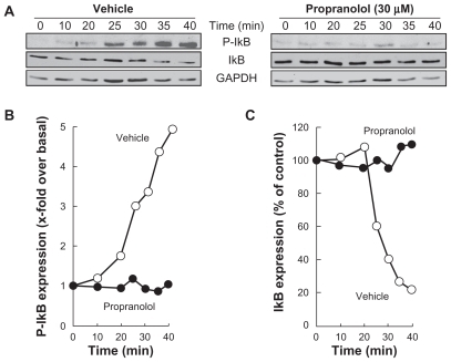
Figure 5 Propranolol inhibits PMA-induced phosphorylation of Erk, and potentiates PMA-mediated Akt phosphorylation. A) Medulloblastoma-derived DAOY cells were serum-starved for 30 minutes in the presence of vehicle or 30 μM propranolol. Cells were then incubated for the indicated time with vehicle or 1 μM PMA. Lysates were isolated, electrophoresed via sodium dodecylsulfate–polyacrylamide gel electrophoresis and immunodetection of phosphorylated Erk (P-Erk), Erk, phosphorylated Akt (P-Akt), and of Akt proteins was performed as described in the Methods section. B, C) Quantification was performed by scanning densitometry of the autoradiograms. Data were expressed as x-fold induction over basal untreated cells for P-Erk/Erk, and as the percent (%) expression of untreated basal conditions for P-Akt/Akt.
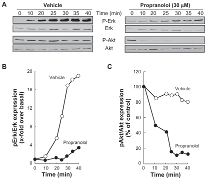
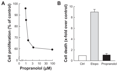
Figure 6 Propranolol inhibits PMA-induced cell proliferation but not cell survival. A) Medulloblastoma-derived DAOY cells were treated as described in and left to grow for 48 hours. Cell proliferation assay was performed as described in the Methods section. B) Cell death was assessed through the release of LDH into the conditioned media and assessed as described in the Methods section of serum-starved DAOY cells treated with vehicle (white bar), 50 μM etoposide (grey bar), or 100 μM propranolol (black bar).