
Figures & data
Table 1 Surgical histopathologic characters
Table 2 Sensitivity, specificity, and positive and negative predictive values for diagnosis of the depth of myometrial invasion with T2WI and T2WI-DWI
Figure 1 Magnetic resonance images of an endometrial cancer patient (stage IA).
Notes: (A) T2-weighted image shows irregular endometrial thickening and appears as heterogeneous signals. (B) DW image shows a lesion with a high signal. (C) Fused T2WI-DWI image shows clear anatomical structures and the lesion shows good contrast from the surrounding normal tissue.
Abbreviations: DWI, diffusion-weighted imaging; T2WI, T2-weighted imaging.
Abbreviations: DWI, diffusion-weighted imaging; T2WI, T2-weighted imaging.
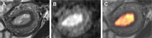
Figure 2 A 56-year-old woman with endometrial carcinoma (pathologic stage is IA).
Notes: (A) T2-weighted image shows that the myometrial signal was heterogeneous (arrow), and we judged the depth of myometrial invasion to be greater than one-half of the myometrial thickness, classifying this case stage as stage IB using T2WI. (B) DW image shows that the extent of the lesion was limited. (C) Fused T2WI-DWI image shows clear margins of the tumor and depth of invasion to be less than one-half of the myometrial thickness.
Abbreviations: DWI, diffusion-weighted imaging; T2WI, T2-weighted imaging.
Abbreviations: DWI, diffusion-weighted imaging; T2WI, T2-weighted imaging.
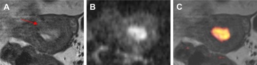
Figure 3 A 60-year-old woman with endometrial carcinoma (pathologic stage, IB).
Notes: (A) It was difficult to judge the depth of myometrial invasion with the T2-weighted image. (B) DW image shows the great extent of the high signal lesion. (C) Fused T2WI-DWI image shows clear margins of the tumor, and the depth of myometrial invasion to be greater than one-half of the myometrial thickness; the diagnosis was clearly stage IB.
Abbreviations: DWI, diffusion-weighted imaging; T2WI, T2-weighted imaging.
Abbreviations: DWI, diffusion-weighted imaging; T2WI, T2-weighted imaging.
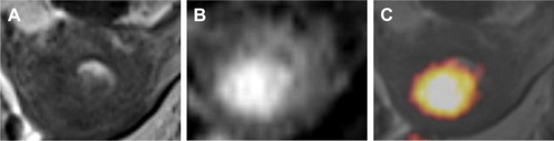
Table 3 Endometrial carcinoma staging with T2WI and fused T2WI-DWI images compared with pathologic results
Table 4 Diagnostic accuracy rate of endometrial carcinoma staging with T2WI and T2WI-DWI