Figures & data
Figure 1 Schematic representation of enzymatic activity and linear structure of human LSD1 and LSD2.
Notes: (A) Enzymatic reaction catalyzed by LSD1. LSD1 catalyzes the demethylation of Lys4 of histone H3 (H3K4) through a flavin-dependent oxidative reaction. LSD1 acts on both di- and monomethylated H3K4. The reaction involves the steps shown from the left to right: first, the histone substrate is bound by the enzyme and the methylated Lys4 side chain is oxidized by the FAD prosthetic group, with consequent reduction of oxygen (O2) to hydrogen peroxide (H2O2); second, the resulting imine intermediate is then hydrolyzed, thus generating the demethylated H3 tail and formaldehyde. (B) Overview of LSD1 and LSD2 structure. Human LSD1 is composed of 852 amino acids with major domains: an N-terminal SWIRM (small α-helical domain), contributing to the steadiness of the molecule; a central protruding Tower domain; and a C-terminal amine oxidase like (AOL) domain. Among these domains, the AOL and SWIRM domains pack together through various interactions (at the level of three-dimensional structure), determining the formation of a spherical structure. At the level of the N-terminal (1–172), there is an N-flexible region. Human LSD2 is composed of 822 amino acids and displays 31% of sequence similarity with LSD1. LSD2 shows three major domains, from the N- to the C-terminal: a ZFs area, a SWIRM, and an AOL domain. It is important to note that LSD2 does not contain a Tower domain, the structure of which plays a crucial role in LSD1 for CoREST binding.
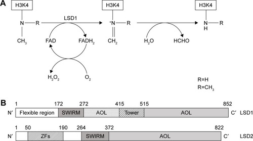
Table 1 Main LSD1 inhibitors introduced into a plan of clinical development
Figure 2 Schematic representation of the DOT1L protein.
Notes: Some physiologically relevant domains are indicated. A large catalytic region (1–332) contains three domains: N′ (1–126), loop (127–140), and an open α/β domain. The N-terminal domain consist of five α-helices and two β-strand hairpins. Loop 127–140 is a protein-flexible loop. The open α/β domain (amino-acid residues 141–332) contains a seven-strand central β-sheet and five α-helices. Part of the flexible loop region and the open α/β domain contributes to the formation of an S-adenosyl-l-methionine (SAM)-binding pocket. A ubiquitin-interaction motif (UIM) is located at amino-acid residues 360–380 and involved in ubiquitin H2B and DOT1L-Bat 3 interactions, both required to facilitate efficient H3K79 dimethylation and trimethylation. A lysine-rich region (380–428) is required for nucleosome binding and interacts with the ubiquitin H2B: deletion of this region resulted in reduced demethylation and trimethylation. Within the lysine-rich region, there is a nucleosome/DNA-binding motif (390–407). Two AF9-binding regions have been mapped: the 628–653 binding region contains one binding site for AF9; the 863–900 binding region contains two AF9-binding sites. The DOT1L865–874-binding domain is strictly required for the binding of the AF9-ENL complex through the C-terminal region of ENL. The 580–1,183 region contains an STT1-binding site.
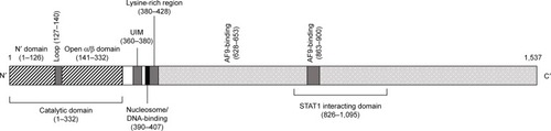
Table 2 Main properties of DOT1L inhibitors
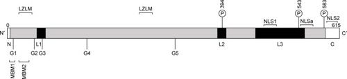
Figure 3 Linear structure of human menin.
Notes: Schematics of a sequence of human menin composed of 615 amino acids. Elements of the folded core of the protein are in gray. These elements contribute to the formation of a central cavity. Three loops, L1–L3, are shown in black. Three phosphorylation sites, located at Ser 394, Ser 543, and Ser 583, are reported and indicated by the symbol P. Two leucine zipper-like motifs (LZLMs) are indicated. Five GTPase motifs (G1–G5) are present in the protein. Two nuclear localization signals (NLS1 and NLS2) and an accessory NLS (NLSa) are reported. The protein possesses two Menin-binding motifs: a high-affinity MBM1 and a low-affinity MBM2.