Figures & data
Figure 1 Immunohistochemical detection of vascular B7-H3 and Tie-2 expression in ccRCC (×400). (A) Vascular distribution (CD34 labeling) in adjacent normal renal tissues showing that renal corpuscle have abundant blood vessels and that blood vessels are scattered in the renal parenchyma. (B) B7-H3 expression was observed in the renal tubular epithelia of adjacent normal tissues, but no expression was detected in glomerular blood vessels and renal parenchymal vessels. (C) Tie-2 expression was found in the renal tubular epithelium and glomerular vascular epithelium of adjacent normal tissues. (D) Low vascular B7-H3 expression in ccRCC tissues. (E) High vascular B7-H3 expression in ccRCC tissues. (F) Low vascular Tie-2 expression in ccRCC tissues. (G) High vascular Tie-2 expression in ccRCC tissues. (H) CD34-labeled MVD in ccRCC tissues. Scale bar 40 µm.
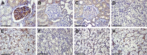
Table 1 Vascular B7-H3 and Tie-2 expressions in ccRCC and correlations with clinicopathological parameters
Table 2 Correlation between B7-H3 and Tie-2 expression in ccRCC
Table 3 MVD in ccRCC and correlations with clinicopathological parameters
Table 4 Relationship between B7-H3, Tie-2 and MVD (mean ± SD) in ccRCC
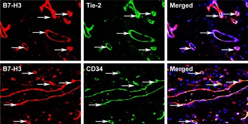
Figure 2 Immunofluorescence staining and laser scanning confocal microscopy revealed B7-H3 (red), Tie-2 (green), and CD34 (green) expression in vascular endothelial cells of ccRCC (arrows). Coexpression of B7-H3/Tie-2 and B7-H3/CD34 was also observed in ccRCC vascular endothelia (arrows).