
Figures & data
Table 1 The basic characteristics of patient with hemorrhage
Figure 1 (A) Crescent-shaped hematoma around the spleen as detected by sonography (arrowheads). (B) Enhanced hemorrhage of puncture tract on contrast-enhanced ultrasound (CEUS). (C) Ultrasound (US)-guided microwave electrode was set into the hemorrhage region (arrowhead). (D) After ablation, enhancement was not detected in the ablation zone (arrowheads).
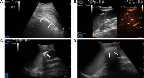
Figure 2 (A and B) Active bleeding originated from the puncture tract with a velocity of 1.13 m/s (arrowheads). (C) A 1.3 cm crescent-shaped free fluid was detected by sonography around the liver (arrowheads). (D) After microwave ablation, contrast-enhanced sonography did not show any microbubble extravasation inside the liver (arrowheads).
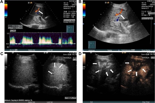
Figure 3 (A) Active bleeding lesion was detected along with small bleeding vessels leaking to the thyroid surface (arrowhead). (B) Contrast-enhanced sonography showed an irregular hematoma measuring 2.6×1.6 cm around the right thyroid (arrowheads). (C and D) Irregular hematoma was detected around the right thyroid, and ablation region showed no enhancement on contrast-enhanced sonography (arrowheads).
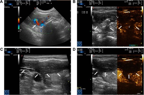
Figure 4 (A and B) Active bleeding originated from a branch of thoracic wall artery (arrowheads). (C) Ultrasound (US)-guided microwave ablation (MWA) (arrowhead). (D) Visible microbubbles extravasation was not observed in this section of hemostasis.
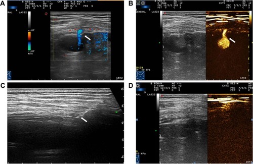
Table 2 The detailed description in the procedures of microwave ablation (MWA) for hemostasis
Table 3 Summary of 12 published literatures about hemostasis in animal and clinical experiments