Figures & data
Figure 1 Ultrasound of the right axillary mass.
Notes: A low echo mass of 3.9×2.9 cm can be seen in the right axillary mammary gland. The boundary is still clear and the shape is irregular. A strong blood flow signal can be detected within it.
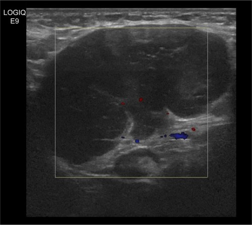
Figure 2 Image of core needle biopsy histologic diagnosis using hematoxylin and eosin staining (original ×100).
Note: Metastasis of poorly differentiated carcinoma can be seen in the resected right axillary lymph node tissue.
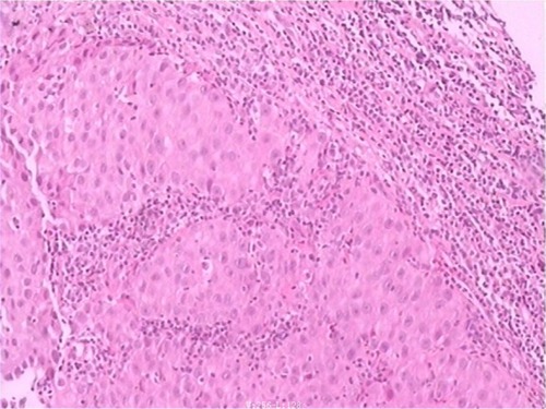
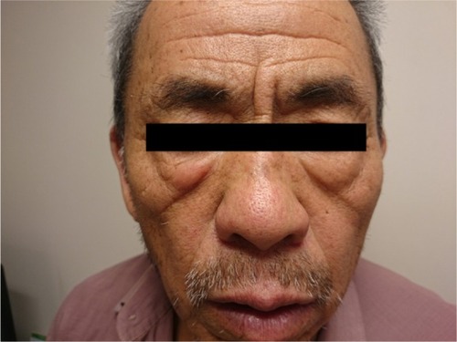
Figure 3 Preoperative facial photo of patient.
Note: Before surgery, the dermal erythematous papules on the right side of the lower eyelid are prominent.
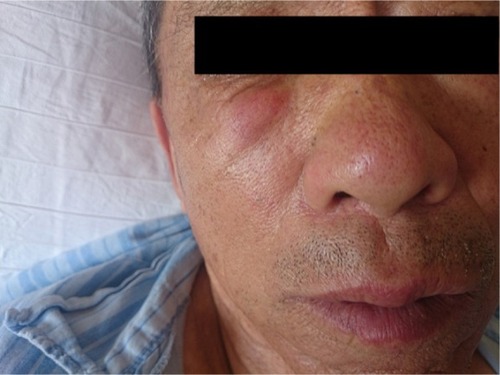
Figure 4 Expression of ER/PR by IHC staining (original ×100) and HER2 by FISH analysis (original ×400).
Notes: (A) ER-negative expression can be detected by IHC staining. (B) PR-negative expression can be detected by IHC staining. (C) HER2-negative expression result can be detected by FISH analysis.
Abbreviations: ER, estrogen receptor; PR, progesterone receptor; IHC, immunohistochemistry; HER2, human epidermal growth factor receptor 2; FISH, fluorescent in situ hybridization.
Abbreviations: ER, estrogen receptor; PR, progesterone receptor; IHC, immunohistochemistry; HER2, human epidermal growth factor receptor 2; FISH, fluorescent in situ hybridization.

Figure 5 Postoperative facial photo of patient.
Note: Ten days after surgery, the dermal erythematous papules on the right side of the lower eyelid have nearly resolved.