Figures & data
Figure 1 Icotinib hydrochloride enhances cisplatin- or radiation-induced proliferation inhibition in Hela S3 cells.
Abbreviations: Ctrl, control; IH, icotinib hydrochloride; IR, ionizing radiation; Cis, cisplatin; CCK-8, Cell Counting Kit-8; h, hours.
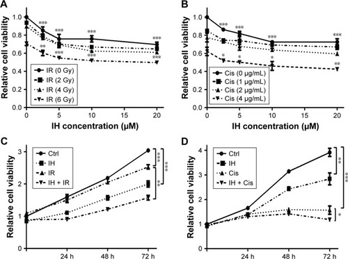
Figure 2 Effect of icotinib hydrochloride on irradiation- or cisplatin-induced cell cycle arrest.
Abbreviations: Ctrl, control; IH, icotinib hydrochloride; IR, ionizing radiation; Cis, cisplatin.
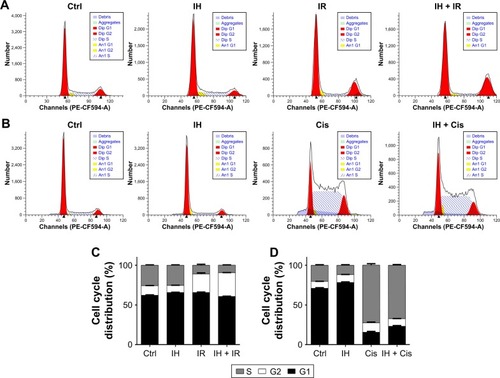
Figure 3 Effect of icotinib hydrochloride on cell apoptosis following radiation or cisplatin treatment.
Abbreviations: Ctrl, control; IH, icotinib hydrochloride; IR, ionizing radiation; Cis, cisplatin.
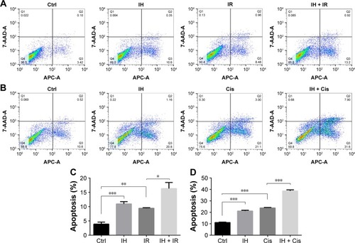
Figure 4 Icotinib hydrochloride inhibits EGFR signaling pathway in Hela S3 cells.
Abbreviations: Ctrl, control; IH, icotinib hydrochloride; IR, ionizing radiation; Cis, cisplatin.
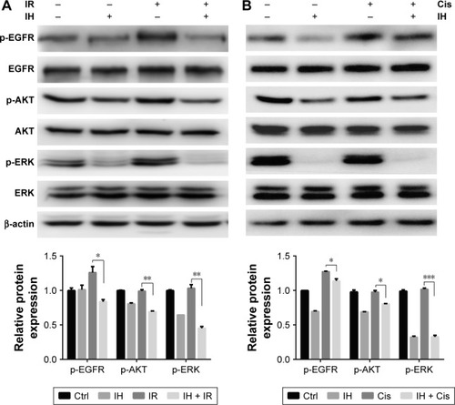
Figure 5 Radiation and chemotherapy sensitization of icotinib hydrochloride by delaying DNA damage repair progress.
Abbreviations: DMSO, dimethyl sulfoxide; IH, icotinib hydrochloride; IR, ionizing radiation; Cis, cisplatin.
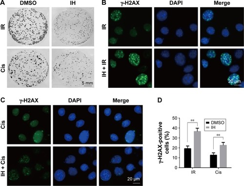
Figure 6 Icotinib hydrochloride attenuates chemo- or radiotherapy-induced HR protein RAD51 upregulation and nuclear foci formation.
Abbreviations: DMSO, dimethyl sulfoxide; qRT-PCR, quantitative reverse transcription polymerase chain reaction; HR, homologous recombination; h, hours; min, minutes; ns, non-significant; Ctrl, control; IH, icotinib hydrochloride; IR, ionizing radiation; Cis, cisplatin; C, cytoplasmic fraction; N, nuclear fraction.
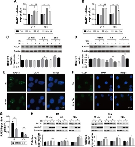
Figure S1 Chemo- and radiotherapy sensitization effects and mechanism of icotinib hydrochloride against SiHa cells.
Abbreviations: DMSO, dimethyl sulfoxide; Ctrl, control; IH, icotinib hydrochloride; IR, ionizing radiation; Cis, cisplatin; C, cytoplasmic fraction; N, nuclear fraction; h, hours.
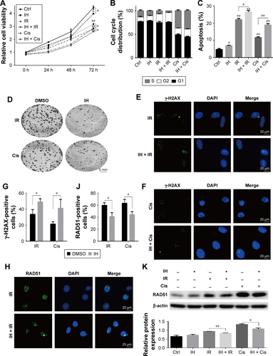
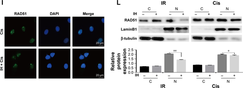
Figure S2 Comparison of EGFR expression levels between Hela S3 and SiHa cells Notes: Results from qRT-PCR (A) and Western blotting (B) assays showing EGFR mRNA levels (A) and protein levels (B) in Hela S3 and SiHa cells. Error bars, SD. **P<0.01 by two-tailed Student’s t-test. All experiments were performed in triplicate.
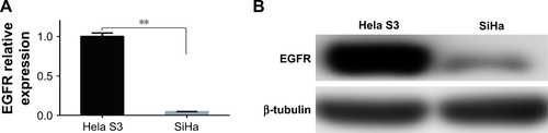
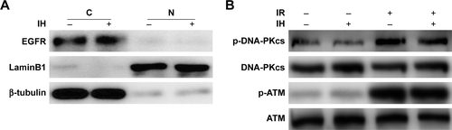
Figure S3 Effects of IH on irradiation-induced nuclear translocation of EGFR and phosphorylation of DNA-PKcs and ATM.
Abbreviations: IH, icotinib hydrochloride; IR, ionizing radiation; C, cytoplasmic fraction; N, nuclear fraction.