Figures & data
Figure 1 Bronchofibroscopic biopsy revealed SCC with opening of right upper lobar bronchus (A), and TBNA found cancer cells in 4R (B) and 7 lymph node groups (C).
Abbreviations: SCC, squamous cell carcinoma; TBNA, transbronchial needle aspiration.
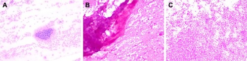
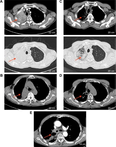
Figure 2 Chest CT images.
Notes: (A and B) the images shows after radiotheray and icotinib, that the right lung primary lesion and mediastinal lymph node were enlarged, and the disease had progressed in March 2016; (C) the image shows tumor shrinkage in May 2016; (D) the image reveals a large cavity in the lung tumor and minimal change in tumor size, in July 2016; (E) the image shows a mass in the right lung hilum and mediastinum, and disease progression once more in October 2017.
Abbreviation: CT, computed tomography.
Abbreviation: CT, computed tomography.