
Figures & data
Table 1 Patient characteristics
Figure 1 Delineation of the heart, pericardium, and LVM.
Notes: (A) Delineation of the heart, pericardium, and LVM in transverse section. (B) Delineation of the heart, pericardium, and LVM in sagittal section. (C) Delineation of the heart, pericardium, and LVM in coronal section. Heart delineation is shown in yellow, pericardium delineation is shown in red, and LVM delineation is shown in blue.
Abbreviation: LVM, left ventricular myocardium.
Abbreviation: LVM, left ventricular myocardium.
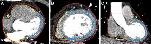
Table 2 Displacements of the heart, pericardium, and LVM in reference to 0% phase CT images (mean ± standard deviation, mm)
Table 3 Volumetric and DSC variations of the heart, pericardium, and LVM
Figure 2 Morphological variations of different phases of the heart, pericardium, and LVM.
Notes: (A) Delineation of the heart in different phases. (B) Morphological variations of different phases of the heart on CT (a significant difference was observed for the left boundary of the LVM). (C) Differences in the left boundary of the LVM are represented using pseudocolors, and significant differences are shown in red. (D) Delineation of the pericardium in different phases. (E) A case showing the LVM in the maximum DSC phase. (F) The same case showing the LVM in the minimum DSC phase. (G) The same case showing the LVM in the maximum and minimum DSC phases in one CT image; the variation of DSC was 700%. (H) Delineation of the LVM in different phases.
Abbreviations: LVM, left ventricular myocardium; CT, computed tomography; DSC, dice similarity coefficient.
Abbreviations: LVM, left ventricular myocardium; CT, computed tomography; DSC, dice similarity coefficient.
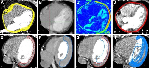
Figure 3 Volumetric variation of the heart, pericardium, and LVM.
Note: The volumetric variation of the LVM was not consistent with those of the heart and pericardium; the volume of heart and pericardium decreased at first and later increased, and the volume of the LVM increased at first and later decreased.
Abbreviation: LVM, left ventricular myocardium.
Abbreviation: LVM, left ventricular myocardium.
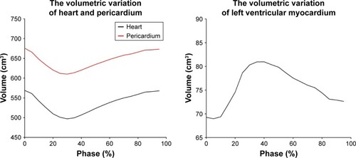
Table 4 Comparison of the morphological parameters of the heart, pericardium, and LVM in the maximum and minimum DSC phases