Figures & data
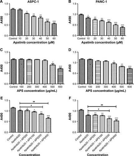
Figure 1 Expression of VEGFR-2 in ASPC-1 and PANC-1 cells and APS enhanced the inhibitory effects of apatinib on cell proliferation.
Abbreviations: APS, Astragalus polysaccharide; VEGFR-2, vascular endothelial growth factor receptor-2; MTS, 3-(4,5-dimethylthiazol-2-yl)-5-(3-carboxymethoxyphenyl)-2-(4-sulfophenyl)-2H-tetrazolium, inner salt; HUVEC, human umbilical vein endothelial cell.
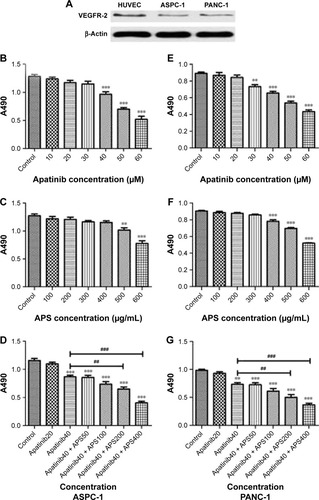
Figure 2 APS enhanced the inhibition of cell migration suppressed by apatinib.
Abbreviation: APS, Astragalus polysaccharide.
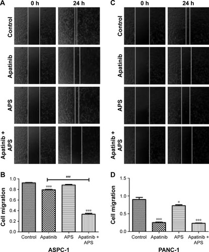
Figure 3 APS enhanced the inhibition of cell invasion suppressed by apatinib.
Abbreviations: AP, apatinib; APS, Astragalus polysaccharide; MMP-9, matrix metalloproteinases-9.
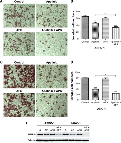
Figure 4 APS increased apoptosis induced by apatinib in pancreatic cancer cells.
Abbreviations: AP, apatinib; APS, Astragalus polysaccharide; FITC, fluorescein isothiocyanate; PI, propidium iodide.
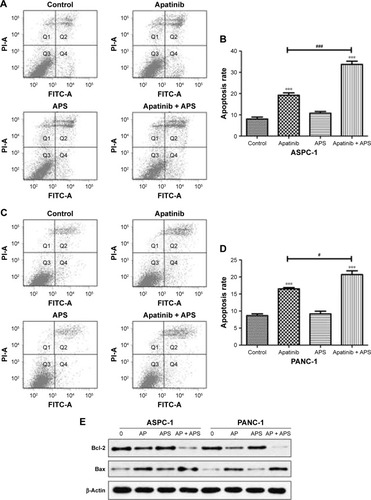
Figure 5 APS increased the inhibition of p-AKT and p-ERK expression in AKT and ERK signaling pathway.
Abbreviations: AP, apatinib; APS, Astragalus polysaccharide; p-AKT, phosphorylated AKT; p-ERK, phosphorylated ERK; AKT, protein kinase B; ERK, extracellular signal-regulated kinase.
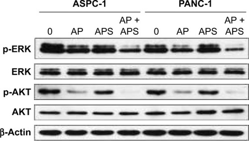
Figure 6 Apatinib and APS induced cellular autophagy.
Abbreviations: AP, apatinib; APS, Astragalus polysaccharide; LC3, light chain 3.
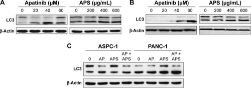
Figure S1 Expression of VEGFR-2 in SW1990 cell line and the influence of apatinib and APS on cell proliferation.
Abbreviations: APS, Astragalus polysaccharide; VEGFR-2, vascular endothelial growth factor receptor-2; MTS, 3-(4,5-dimethylthiazol-2-yl)-5-(3-carboxymethoxyphenyl)-2-(4-sulfophenyl)-2H-tetrazolium, inner salt.
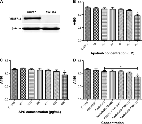
Figure S2 Apatinib inhibited cell proliferation more significantly after adding rhVEGF-165 in ASPC-1 and PANC-1.
Abbreviations: APS, Astragalus polysaccharide; MTS, 3-(4,5-dimethylthiazol-2-yl)-5-(3-carboxymethoxyphenyl)-2-(4-sulfophenyl)-2H-tetrazolium, inner salt.