Figures & data
Figure 1 Imaging for Case 1.
Notes: Pre-treatment (A, B) and post-treatment (C, D) fused axial PET/CT images of the patient’s left-sided primary breast lymphoma (A, C) and gastrointestinal stromal tumor of the pancreatic head (B, D). Pre-treatment imaging revealed mildly FDG-avid left breast changes at the site of biopsy-confirmed DLBCL (A), with resolution of these findings after six cycles of R-CHOP chemotherapy (C). Pre-treatment imaging revealed a 5.0 × 6.0 cm duodenal/pancreatic head mass with significant FDG avidity (B); this decreased in size to 4.6 × 5.2 cm after 10 months of imatinib, with continued FDG avidity, suggesting persistent disease despite partial response (D).
Abbreviations: DLBCL, diffuse large B cell lymphoma; FDG, fluorodeoxyglucose; PET, positron emission tomography; R-CHOP, rituximab/cyclophosphamide/doxorubicin/vincristine/prednisolone.
Abbreviations: DLBCL, diffuse large B cell lymphoma; FDG, fluorodeoxyglucose; PET, positron emission tomography; R-CHOP, rituximab/cyclophosphamide/doxorubicin/vincristine/prednisolone.
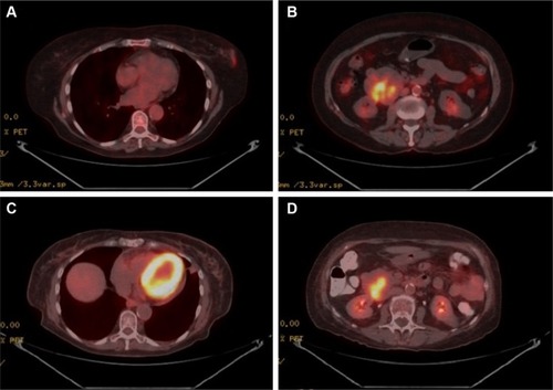
Figure 2 Case 1 GIST surgical resection pathology findings.
Notes: (A) Gastrointestinal stromal tumor involving the wall of the duodenum (arrowhead). The neoplastic cells showed epithelioid morphology with focal areas of spindle cell morphology. Mitotic figures were few (<5 per 50 high-power fields). (H&E stain; 100× original magnification) (B) DOG1 immunohistochemistry showed diffuse positive staining. (C) Bcl-2 immunohistochemistry showed diffuse positive staining. (Immunohistochemistry with hematoxylin counterstain; 100× original magnification).
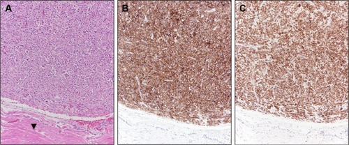
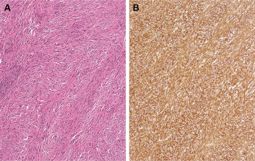
Figure 3 Case 2 GIST surgical resection pathology findings.
Notes: (A) GIST comprised of neoplastic cells with spindle cell morphology exhibiting a herringbone growth pattern. Mitotic figures were few (<5 per 50 high-power fields). (H&E stain; 100× original magnification) (B) CD117 immunohistochemistry showed diffuse positive staining. (Immunohistochemistry with hematoxylin counterstain; 100× original magnification).
Abbreviation: GIST, gastrointestinal stromal tumor.
Abbreviation: GIST, gastrointestinal stromal tumor.