Figures & data
Table 1 Clinical characteristics of patients with different stages of gastric carcinogenesis
Figure 1 PLK1 is upregulated in gastric cancer tissues. Expression level of PLK1 was measured in gastric tumor samples (T) and their non-cancerous counterparts (N). GAPDHwas used as an internal control. Densitometric units of T are expressed as a fold of its corresponding N. ***p<0.001.
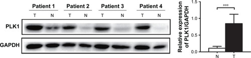
Figure 2 Expression of PLK1 in different stages of gastric carcinogenesis in relation to Helicobacter pylori. Gastric tissue samples were stained with antibodies against PLK1; (A) immunoreactive cells positive for PLK1 were semi quantitatively assessed and the protein expression levels are expressed as grade 1–4; (B) all patients in various stages of gastric lesions; (C, D) patients with various stages of gastric lesions with or without H. pylori infection. Mean grades () for protein expression are shown. *p<0.05; **p<0.01; ***p<0.001.
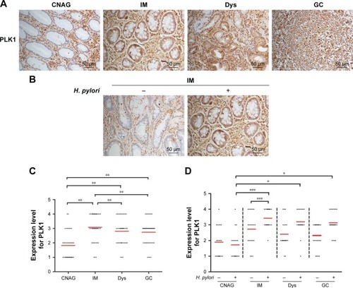
Figure 3 Helicobacter pylori infection induces high expression of PLK1 in gastric tissue from Mongolian gerbils. (A) Gastric tissue sections from H. pylori-infected gerbils were stained with antibodies against PLK1. (B) Immunoreactive cells were semi quantitatively assessed and the protein expression levels are expressed as grade 1–4. Mean grades () for protein expression are shown. *p<0.05; **p<0.01; ***p<0.001.
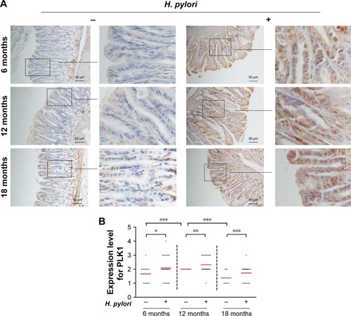
Figure 4 PLK1 is involved in Helicobacter pylori-promoted cancer cell proliferation. Cell survival was evaluated by a CCK-8 assay and the cell growth rate was expressed as a percentage of the control or MKN-28 cells at various time points after (A) incubation with different doses of H. pylori (MOI = 10, 50, 100, 200) and (B) pretreatment with an inhibitor of PLK1 BI 2536. The data are representative of three independent experiments. *p<0.05; ***p<0.001.
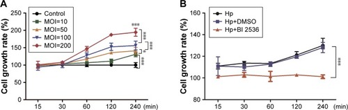
Figure 5 Helicobacter pylori infection induces PLK1-mediated activation of the PI3K/Akt pathway in a dose-dependent manner. Cultures of MKN-28 cells were incubated with various doses of H. pylori (at 2 h) in the H. pylori group or with medium in control group. Immunoblotting was used to quantify the expression levels of PLK1 (A) and Akt pathway-related proteins (B). The data are representative of three independent experiments. The samples were derived from the same experiment, and the blots were processed in parallel. *p<0.05; **p<0.01; ***p<0.001.
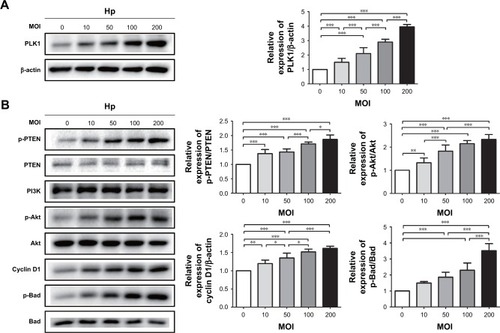
Figure 6 Helicobacter pylori infection induces PLK1-mediated activation of the PI3K/Akt pathway in a time-dependent manner. Cultures of MKN-28 cells were incubated at different time points with H. pylori infection (MOI = 200). Immunoblotting was used to quantify the expression levels of PLK1 (A) and Akt pathway-related proteins (B). The data are representative of three independent experiments. The samples were derived from the same experiment and the blots were processed in parallel. **p<0.01; ***p<0.001.
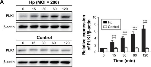
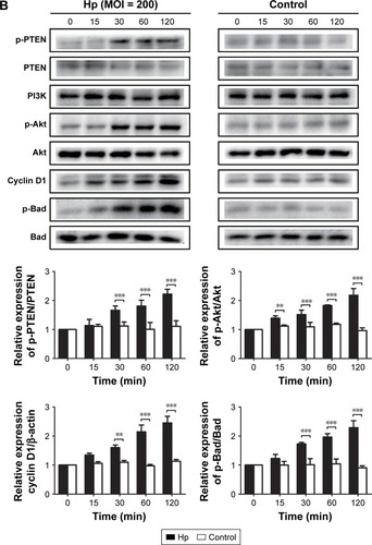
Figure 7 PLK1 inhibition blocks Helicobacter pylori-induced activation of the Akt pathway. Immunoblotting was used to quantify the expression levels of PLK1 and Akt pathway-related proteins in MKN-28 cells incubated with H. pylori for 24 h and treated with BI 2536 (200 nM pretreatment before incubation with H. pylori). The data are representative of three independent experiments. The samples were derived from the same experiment and the blots were processed in parallel. **p<0.01; ***p<0.001.
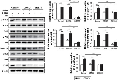
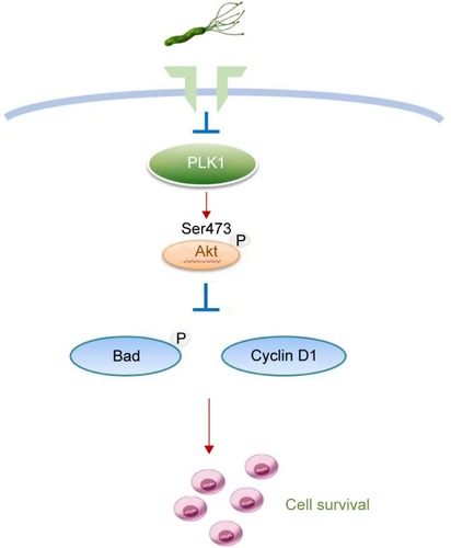
Figure 8 A schematic model of GC development. Helicobacter pylori promotes gastric epithelial cell survival through the PLK1/PI3K/Akt pathway.