Figures & data
Figure 1 Abdominal ultrasound showed a 2.9×2.6 cm2 hypoechoic solid tumor with abundant blood flow signal.
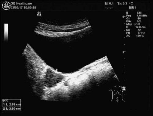
Figure 2 Pelvic magnetic resonance imaging showed that a 3.1×2.6 cm2 nodule (blue arrows) had invaded the rectum.
Notes: (A) T1 weighted image with sagittal view; (B) T2 weighted image with sagittal view; (C) T1 weighted image with axial view; (D) T2 weighted image with axial view.
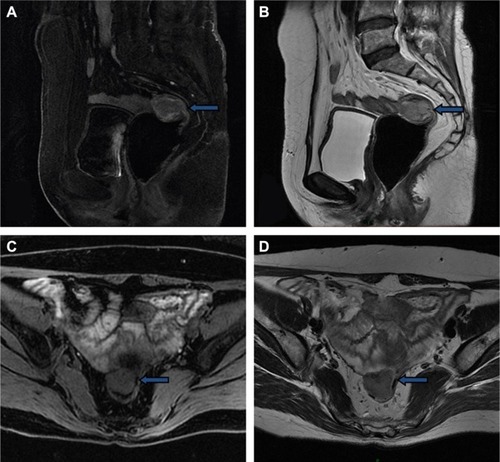
Figure 3 Trocar placement and the size of the trocars.
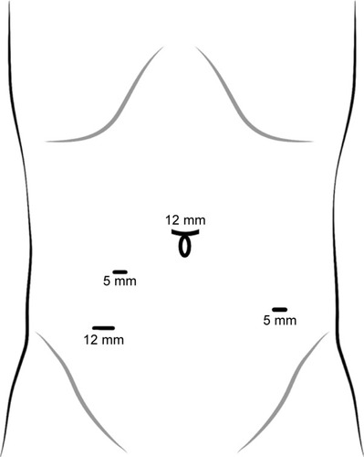
Figure 4 Transanal specimen extraction via laparoscopic rectectomy without an abdominal incision.
Notes: (A) The tumor was located at the right front wall of the middle rectum; (B) naked intestines at 3 cm from the distal margin of the tumor; (C) naked intestines at 3 cm from the proximal margin of the tumor; (D) the distal rectum was dissected circularly; (E) the transected bowel was pulled out via the anus; (F) the distal circular stapling device anvil was fixed extracorporeally; (G) the colon was then repositioned into the abdomen; (H) the rectal stump was closed; (I) an endtoend circular anastomosis was performed.
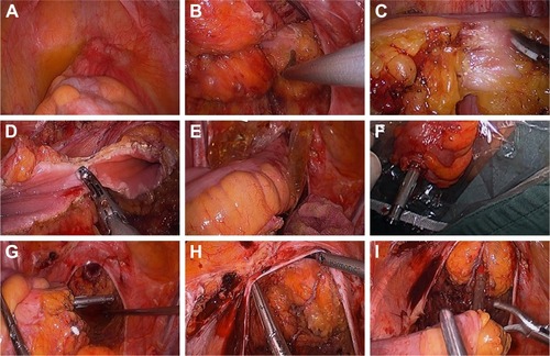
Figure 5 Macroscopic observation of rectal neoplasm.
Notes: (A) Rectal serous membrane. (B) Rectal mucosal membrane.
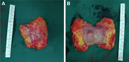
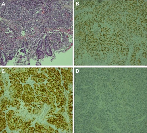
Figure 6 Microscopic observation and immunochemistry of rectal neoplasm.
Notes: (A) Microphotography shows poorly differentiated cells of adenocarcinoma arranged in nests, with vessel invasion (HematoxylinEosin G×100); (B) the immunochemistry showed that cells were WT1(3+); (C) the immunochemistry showed that cells were PAX2(3+); (D) the immunochemistry showed that cells were CDX2(−).