Figures & data
Figure 1 The expression of DcR3 in gastric cancer cell lines and generation of DcR3 knockdown cell lines.
Notes: (A) The expression of DcR3 in different GC cells was measured by enzyme-linked immunosorbent assay and quantified by RT-PCR. (B) Images captured by fluorescence microscope showing the fluorescent signal of the enhanced green fluorescent protein in stably transfected cells. (C and D) HGC27 and MKN28 cells of all groups were harvested for DcR3 measurement by Western blotting and RT-PCR. Expression of DcR3 was decreased significantly in LV-SiDcR3 group compared with control group and LV-NC group (P<0.05). ***P<0.001.
Abbreviations: DcR3, decoy receptor 3; GAPDH, glyceraldehyde 3-phosphate dehydrogenase; RT-PCR, reverse transcriptase polymerase chain reaction; LV-NC, lentiviral negative control.
Abbreviations: DcR3, decoy receptor 3; GAPDH, glyceraldehyde 3-phosphate dehydrogenase; RT-PCR, reverse transcriptase polymerase chain reaction; LV-NC, lentiviral negative control.
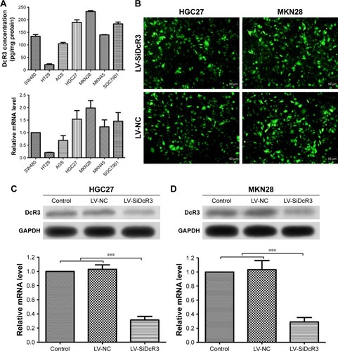
Figure 2 Effects of DcR3 knockdown on proliferation, colony formation, cell cycle distribution and apoptosis of HGC27 and MKN28 cells.
Notes: (A) Colony formation ability and cell growth ability of 3 groups (Control, LV-NC and LV-SiDcR3 cells). (B) Effects of DcR3 inhibition on cell cycle of cells in various groups (Control, LV-NC and LV-SiDcR3). HGC27 LV-SiDcR3 group showed G2/M phase arrest, while MKN28 LV-SiDcR3 group showed G1 and G2/M phase arrest. (C) Cell apoptosis was determined using flow cytometry, and the apoptosis level was enhanced after transfection with siRNA in HGC27 and MKN28 cell lines. All assays were repeated three times. *P<0.05, **P<0.01, and ***P<0.001.
Abbreviations: DcR3, decoy receptor 3; FITC, fluorescein isothiocyanate; LV-NC, lentiviral negative control.
Abbreviations: DcR3, decoy receptor 3; FITC, fluorescein isothiocyanate; LV-NC, lentiviral negative control.
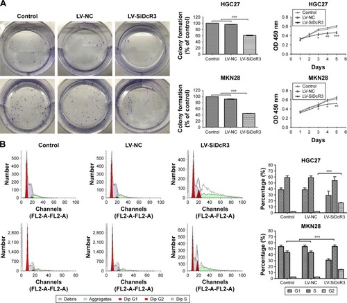
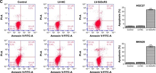
Figure 3 Cell mobility was inhibited after transfection with DcR3 siRNA in HGC27 and MKN28 cell lines.
Notes: (A) Representative set of images showing the migration of HGC27 cells toward scratch wound. Graph presenting the normalized scratch width relative to control group at 0 and 24 hours after the scratch, the pairwise comparisons between LV-SiDcR3 and control and LV-NC groups were statistically significant (P<0.05). (B) Cell invasion was determined using Transwell assay. Cell invasion was significantly inhibited after transfection with siRNA in HGC27 and MKN28 cells. (C) The wound healing assay showed cell migration was inhibited after transfection with siRNA in MKN28 cells. (D) Cell migration was determined using Transwell assay. Cell migration was significantly inhibited after transfection with siRNA in HGC27 and MKN28 cells. (E and F) DcR3 overexpression in HGC27 and MKN28 cells caused morphological alteration resembling EMT, while knockdown of DcR3 attenuated the morphological alteration. ***P<0.001.
Abbreviation: DcR3, decoy receptor 3; LV-NC, lentiviral negative control.
Abbreviation: DcR3, decoy receptor 3; LV-NC, lentiviral negative control.
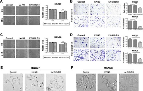
Figure 4 Silencing DcR3 inhibited PI3K/Akt/GSK-3β/β-catenin signaling in HGC27 and MKN28 cells.
Notes: (A and C) The main protein components of the PI3K/Akt/GSK-3β/β-catenin signaling pathway in HGC27 and MKN28 cells were measured by Western blot assay. (B and D) The relative expression of each protein to GAPDH in HCG27 and MKN28 cells. *P<0.05, **P<0.01, and ***P<0.001.
Abbreviations: DcR3, decoy receptor 3; GAPDH, glyceraldehyde 3-phosphate dehydrogenase; LV-NC, lentiviral negative control.
Abbreviations: DcR3, decoy receptor 3; GAPDH, glyceraldehyde 3-phosphate dehydrogenase; LV-NC, lentiviral negative control.
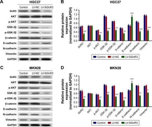
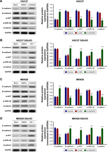
Figure 5 Effects of LY294002 and IGF-1 on HGC27 and MKN28 cells.
Notes: (A and C) HGC27 and MKN29 cells were treated with LY294002 (50 µM) for 24 hours, and PI3K/Akt/GSK-3β/β-catenin signaling and downstream molecule expression levels were analyzed by Western blotting. (B and D) HGC27 and MKN28 DcR3 knockdown cells were treated with IGF-1 (100 ng/mL) for 24 hours, and PI3K/Akt/GSK-3β/β-catenin signaling and downstream molecule expression levels were analyzed by Western blotting. **P<0.01 and ***P<0.001.
Abbreviations: DcR3, decoy receptor 3; GAPDH, glyceraldehyde 3-phosphate dehydrogenase; IGF, insulin-like growth factor; LV-NC, lentiviral negative control.
Abbreviations: DcR3, decoy receptor 3; GAPDH, glyceraldehyde 3-phosphate dehydrogenase; IGF, insulin-like growth factor; LV-NC, lentiviral negative control.