Figures & data
Figure 1 CLDN6 expression level in EC was significantly upregulated.
Abbreviations: UCEC, Uterine Corpus Endometrial Carcinoma; ANT, adjacent non-tumorous; CLDN6, claudin-6; EC, endometrial carcinoma; ESC, endometrial cells; TCGA, The Cancer Genome Atlas.

Table 1 Clinical association between CLDN6 expression and clinicopathological variables in EC patients
Table 2 Univariate and multivariate analysis of clinical prognostic factors of EC
Figure 2 Kaplan–Meier OS curves based on CLDN6 expression level.
Abbreviations: CLDN6, claudin-6; EC, endometrial carcinoma; OS, overall survival.

Figure 3 Knockdown efficiency was determined by qRT-PCR (A) and western blotting (B and C) in EC cells.
Abbreviations: CLDN6, claudin-6; EC, endometrial carcinoma.
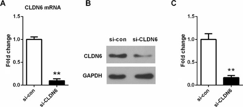
Figure 4 Knockdown CLDN6 in EC cells significantly reduced the proliferative abilities, as determined by CCK-8 assay.
Abbreviations: CCK-8, cell counting kit-8; CLDN6, claudin-6; EC, endometrial carcinoma.
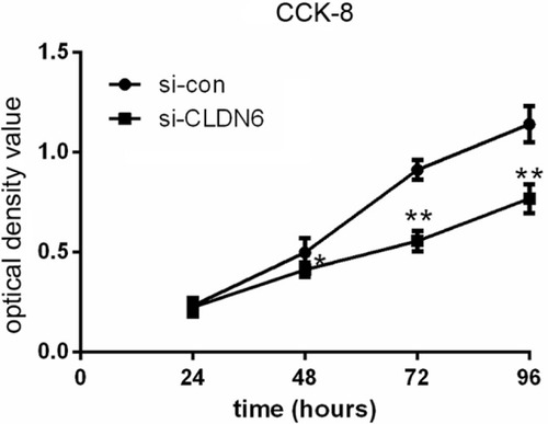
Figure 5 Colony-formation assays suggested that knockdown of CLDN6 significantly reduced the colony-forming ability of EC cells.
Abbreviations: CLDN6, claudin-6; EC, endometrial carcinoma.
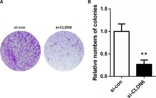
Figure 6 Migratory/invasive capacity was measured using wound-healing and transwell assay.
Abbreviation: CLDN6, claudin-6.
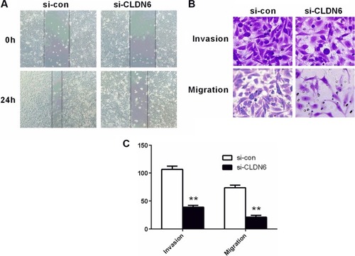
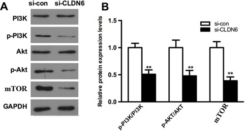
Figure 7 Effect of CLDN6 knockdown on activation of PI3K pathway in HEC-1B cells.
Abbreviations: CLDN6, claudin-6; EC, endometrial carcinoma.