
Figures & data
Table 1 Relationship between CPEB4 expression and clinico-pathological features in gastric cancer
Table 2 Correlation analysis between CPEB4 expression and ZEB1 expression in gastric cancer tissues by chi-squared test
Figure 1 Relative CPEB4 expression in GC tissues and its clinical significance.
Abbreviations: CPEB4, cytoplasmic polyadenylation element-binding protein 4; GC, gastric cancer; IHC, immunohistochemistry.
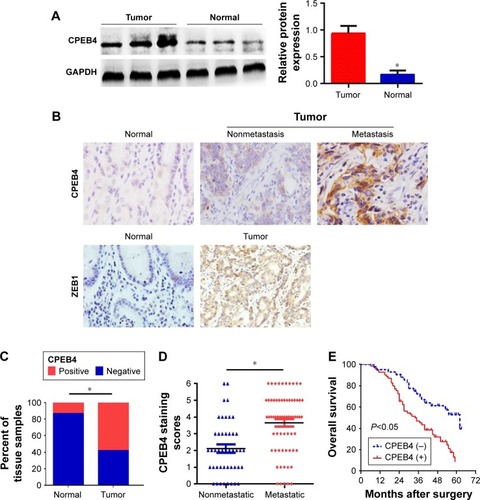
Figure 2 Effects of CPEB4 silencing or overexpression on GC cells’ migration, invasion, and proliferation in vitro.
Abbreviations: CCK-8, cell-counting kit-8; CPEB4, cytoplasmic polyadenylation element-binding protein 4; GC, gastric cancer.
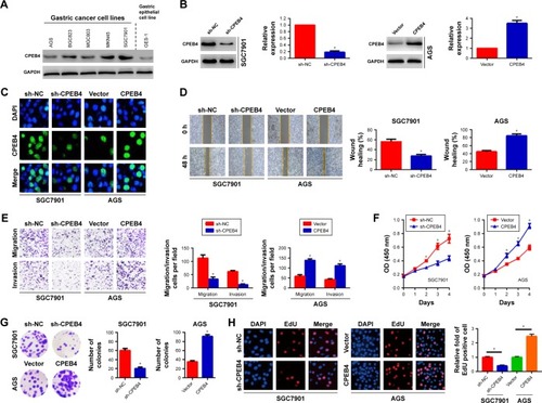
Figure 3 Effects of different expression levels of CPEB4 on EMT-related markers in SGC7901 and AGS cells.
Abbreviations: CPEB4, cytoplasmic polyadenylation element-binding protein 4; EMT, epithelial–mesenchymal transition; qRT-PCR, quantitative real-time PCR; shRNA, short hairpin RNA.
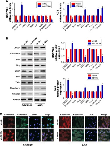
Figure 4 CPEB4 induced GC cells’ migration, invasion, and growth via ZEB1-mediated EMT.
Abbreviations: CCK-8, cell-counting kit-8; CPEB4, cytoplasmic polyadenylation element-binding protein 4; EMT, epithelial–mesenchymal transition; GC, gastric cancer.
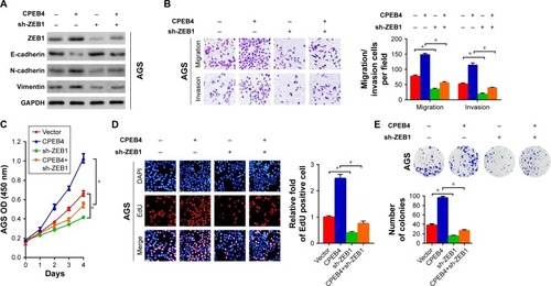
Figure 5 Influences of CPEB4 silencing or overexpression on GC cells’ tumor growth in vivo.
Abbreviations: CPEB4, cytoplasmic polyadenylation element-binding protein 4; GC, gastric cancer.
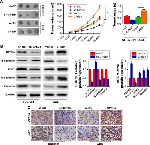
Figure 6 Effects of CPEB4 silencing or overexpression on the metastasis of GC cells in vivo.
Abbreviations: CPEB4, cytoplasmic polyadenylation element-binding protein 4; GC, gastric cancer.
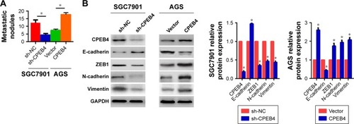
Table S1 Primers designed for qRT-PCR