Figures & data
Figure 1 miR-222-3p is upregulated in OS tissues and cell lines.
Notes: (A) miR-222-3p mRNA levels were examined by qRT-PCR analysis in 30 cases of clinical OS tissues and paired peritumor tissues. **P<0.01. (B) Gene expression levels of miR-222-3p were compared between human osteosarcoma cell lines (HOS, MG-63, Saos-2, and U-2OS) and normal human osteoblast cell hFOB by qRT-PCR. *P<0.05; **P<0.01.
Abbreviations: OS, osteosarcoma; RT-qPCR, reverse-transcription quantitative PCR.
Abbreviations: OS, osteosarcoma; RT-qPCR, reverse-transcription quantitative PCR.
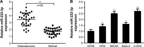
Figure 2 Inhibition of miR-222 in OS cells decreases their proliferation and colony formation.
Notes: (A, B) miR-222-3p inhibitor or NC was transfected into MG-63 and U-2OS cells, respectively, and miR-222-3p expression was measured using RT-qPCR. (C, D) Cell proliferation was assayed in MG-63 and U-2OS cells. Cells transfected with miR-222-3p inhibitor displayed decreased proliferation compared with the control. (E, F) Colony formation was assayed in MG-63 and U-2OS cells. *P<0.05; **P<0.01.
Abbreviations: OS, osteosarcoma; RT-qPCR, reverse-transcription quantitative PCR; NC, control inhibitor.
Abbreviations: OS, osteosarcoma; RT-qPCR, reverse-transcription quantitative PCR; NC, control inhibitor.
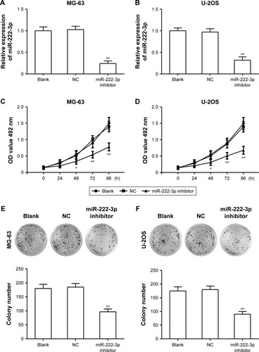
Figure 3 Inhibition of miR-222 suppresses migration and invasion in OS cells.
Notes: (A, B) Cell migration was determined by wound healing assay in MG-63 and U-2OS cells after transfection with miR-222-3p inhibitor or control inhibitor. (C, D) Transwell invasion assay was conducted in MG-63 and U-2OS cells. *P<0.05; **P<0.01.
Abbreviation: OS, osteosarcoma.
Abbreviation: OS, osteosarcoma.
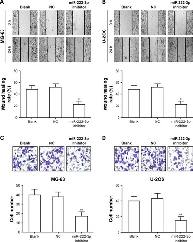
Figure 4 TIMP3 is a direct target of miR-222-3p.
Notes: (A) The suspected binding of mature human miR-222-3p with the wild-type 3′-UTR region of TIMP3 mRNA is shown. A mutated 3′-UTR of TIMP3 is also shown. (B, C) A dual-luciferase reporter assay was performed with MG-63 and U-2OS cells cotransfected with firefly luciferase constructs containing wild-type 3′-UTR region of TIMP3 (Wt-TIMP3) or mutated 3′-UTR region of TIMP3 (Mut-TIMP3) and miR-222-3p mimic or control. The relative luciferase activities were evaluated 24 hours after transfection (**P<0.05). (D, E) Osteosarcoma MG-63 and U-2OS cells were infected by miR-222-3p mimic, miR-222-3p inhibitor, or control. The protein expression levels of TIMP3 were evaluated by Western blotting.
Abbreviations: TIMP3, tissue inhibitor of metalloproteinases 3; UTR, untranslated region; NC, control inhibitor.
Abbreviations: TIMP3, tissue inhibitor of metalloproteinases 3; UTR, untranslated region; NC, control inhibitor.
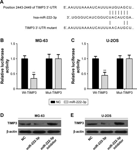
Figure 5 TIMP3 is inversely expressed with miR-222-3p in OS tissues.
Notes: (A) The gene expression levels of TIMP3 were compared between human clinical OS tissues and paired peritumoral tissues (n=30, **P<0.01). (B) Correlation of miR-222-3p levels with TIMP3 mRNA levels was examined by RT-qPCR analysis in clinical OS tissues (Pearson’s correlation coefficient, r=−0.754; n=30; P<0.01).
Abbreviations: TIMP3, tissue inhibitor of metalloproteinases 3; OS, osteosarcoma; RT-qPCR, reverse-transcription quantitative PCR.
Abbreviations: TIMP3, tissue inhibitor of metalloproteinases 3; OS, osteosarcoma; RT-qPCR, reverse-transcription quantitative PCR.
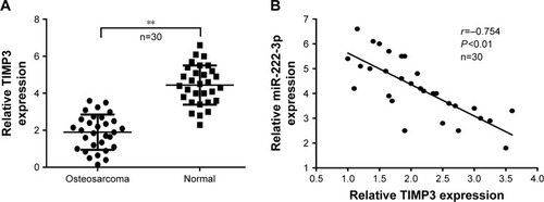
Figure 6 TIMP3 is involved in miR-222-3p-mediated regulation of OS cells.
Notes: (A) The interference efficiency of TIMP3 siRNAs (si-TIMP3-1 or si-TIMP3-2) was examined by Western blotting in hFOB cells. (B) Effects of miR-222-3p inhibitor and miR-222-3p inhibitor+si-TIMP3 on MG-63 cell growth. (C) Effects of miR-222-3p inhibitor and miR-222-3p inhibitor+si-TIMP3 on MG-63 cell colony formation ability. (D) Effects of miR-222-3p inhibitor and miR-222-3p inhibitor+si-TIMP3 on MG-63 cell invasion ability. *P<0.05; **P<0.01.
Abbreviations: TIMP3, tissue inhibitor of metalloproteinases 3; OS, osteosarcoma; NC, control inhibitor.
Abbreviations: TIMP3, tissue inhibitor of metalloproteinases 3; OS, osteosarcoma; NC, control inhibitor.
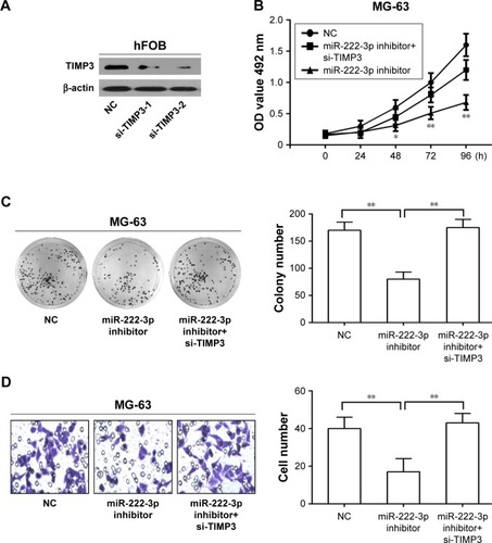
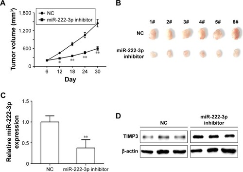
Figure 7 Inhibition of miR-222-3p reduces tumor growth in vivo.
Notes: (A) Tumor growth curves. (B) Photographs of tumor tissues. (C) miR-222-3p expression in tumor tissues was determined by RT-qPCR analysis. (D) TIMP3 protein expression in tumor tissues was determined by Western blotting. β-actin was used as an internal control. *P<0.05; **P<0.01.
Abbreviations: RT-qPCR, reverse transcription-quantitative polymerase chain reaction; TIMP3, tissue inhibitor of metalloproteinases 3; NC, control inhibitor.
Abbreviations: RT-qPCR, reverse transcription-quantitative polymerase chain reaction; TIMP3, tissue inhibitor of metalloproteinases 3; NC, control inhibitor.