Figures & data
Table 1 Criteria for positive staining for in situ hybridization
Table 2 Number of colonies formed by different cell typesTable Footnotea
Figure 1 It was shown by qRT-PCR tests of miR-30c that its expression was significantly different between the normal and osteosarcoma cells (4.62±1.12 vs 1.37±0.42, P<0.01).
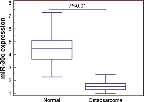
Figure 2 In situ hybridization tests showed that the relative expression level of miR-30c was significantly higher in the bone tissue than in osteosarcoma tissue (3.92±0.94 vs 1.23±0.45, P<0.001, scale bar: 100 µm, magnification: ×200), which is similar to that of qRT-PCR analysis.
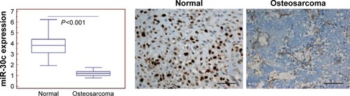
Table 3 Relationship between the clinical characteristics of patients and the expression of miR-30c
Table 4 Low expression level of miR-30c and the prognosis of osteosarcoma patients
Table 5 Multilinear regression analysis of osteosarcoma patient prognosis
Figure 3 Kaplan–Meier analysis of osteosarcoma patients: low expression level of miR-30c is closely related to the short survival of patients (P<0.01).
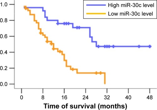
Figure 4 Effect of miR-30c on the colony-forming ability of U2OS cells.
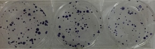
Figure 5 The proliferation capacity of cells was significantly lower in the experimental group than in the control group at 72, 96 and 120 hours after transfection (*P<0.05).
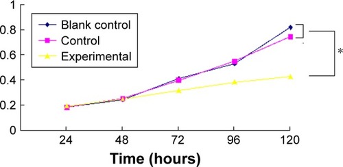
Figure 6 There was no significant difference between the blank control group and the control group (P>0.05, one-way ANOVA), but there were significant differences between the experimental group and other groups (P<0.01, one-way ANOVA).
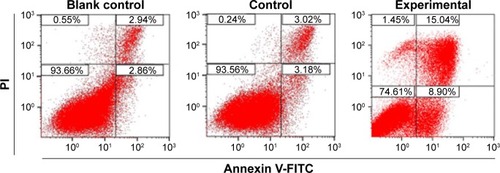
Figure 7 After transfecting U2OS cells with miR-30c mimics, their migration and invasiveness were found to be significantly haltered inhibited.
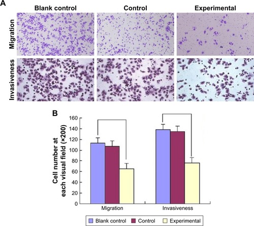
Table 6 Weight and volume of the tumors in the three groups of nude miceTable Footnotea
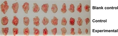
Figure 8 The difference was significant among the groups on comparing the volume of percutaneously formed tumors.