Figures & data
Table 1 Primer sequences for the amplification of target genes
Figure 1 Tumor cells and BM-MSCs induce platelet activation.
Abbreviations: BM-MSC, bone-marrow mesenchymal stem cell; CM, conditioned medium; FITC, fluorescein isothiocyanate; SEM, standard error of the mean; SSC, side scatter.
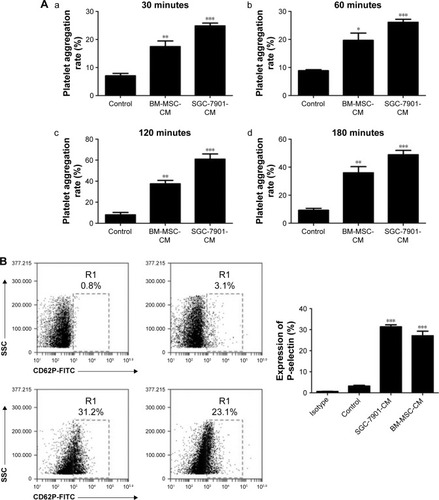
Figure 2 Platelets induce BM-MSCs transdifferentiation into CAF-like cells by secreting TGF-β.
Abbreviations: BM-MSCs, bone-marrow MSCs; CAF, cancer-associated fibroblast; FAP, fibroblast activation protein; GAPDH, glyceraldehyde-3-phosphate dehydrogenase; MSC, mesenchymal stem cell; PLT, platelet; SEM, standard error of the mean; α-SMA, α-smooth muscle actin.
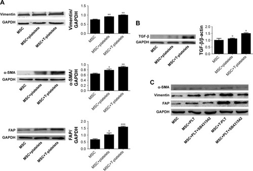
Figure 3 Platelets promote BM-MSCs proliferation and migration.
Abbreviations: MSC, mesenchymal stem cell; PLT, platelet; BM-MSCs, bone-marrow MSCs.
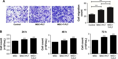
Figure 4 Platelets enhanced the effect of BM-MSCs on proliferation and metastasis of tumor cells.
Abbreviations: CM, conditioned medium; MSC, mesenchymal stem cell; PLT, platelet; VEGF, vascular endothelial growth factor; BM-MSCs, bone-marrow MSCs; SEM, standard error of the mean.
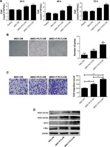
Figure 5 Platelets enhanced the effect of BM-MSCs on tumor progression in vivo.
Abbreviations: CM, conditioned medium; MSC, mesenchymal stem cell; PLT, platelet; VEGF, vascular endothelial growth factor; BM-MSCs, bone-marrow MSCs; SEM, standard error of the mean.
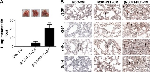
Figure S1 Characteristic surface markers of BM-MSCs were detected by flow cytometry analysis.
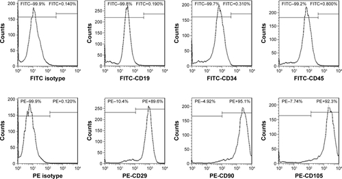
Figure S2 (A) Platelet aggregation of platelets co-cultured with SGC-7901 cells for 0 hours (a, b) and 2 hours (c, d) was photographed by microscope. (B) Platelet aggregation of platelets co-cultured with BM-MSCs for 0 hours (a, b) and 2 hours (c, d) was photographed by microscope. The magnification of a and c is ×400 (scale bar: 10 µm) and the magnification of b and d is ×200 (scale bar: 20 µm).
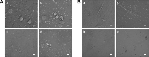
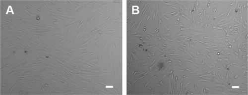
Figure S3 Cell morphology of BM-MSCs co-cultured with T-platelets for 0 hours (A) and 24 hours (B).
Abbreviation: BM-MSCs, bone marrow-derived mesenchymal stem cells.