Figures & data
Figure 1 HIFU, together with bufalin, impairs the growth of PC cells and induces the apoptosis of PC cells.
Notes: The colony formation assay was used to assess the growth of pancreatic cells. HIFU, together with bufalin, inhibited the growth of MiaPaCa2 (A) and Panc-1 (B) cells. Flow cytometric analysis was performed to evaluate the induction of apoptosis in PC cells. The proportion of MiaPaCa2 cells in apoptosis was highest in the HIFU+bufalin-treated group than in the bufalin-treated or HIFU-treated groups (C). An increased number of apoptotic Panc-1 cells was observed after HIFU+bufalin treatment than after treatment with bufalin or HIFU alone (D). The survival rates of cells in the control group, the bufalin group, the HIFU group, and the HIFU+bufalin group were 90.9%, 60.2%, 39.7%, and 17.3% (E), respectively. The survival rates of cells in the control group, the bufalin group, the HIFU group, and the HIFU+bufalin group were 97.5%, 53.9%, 40.9%, and 13.8%, respectively (F). Error bars represent the mean±SEM of three independent experiments. Data shown are representative of three independent experiments. *P<0.05 and ***P<0.0001, one-way ANOVA.
Abbreviations: HIFU, high-intensity focused ultrasound; ns, nonsignificant.
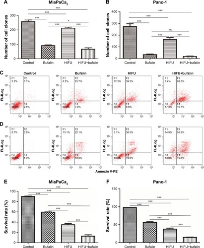
Figure 2 Bufalin, combining HIFU, induces expression of apoptosis-related proteins. PARP, cleaved caspase-3, caspase-8, caspase-9, Bax, and Bak protein expression in (A) MiaPaCa2 cells and (B) Panc-1 cells were examined by Western blotting.
Note: Representative blots from three biological repeat experiments are shown.
Abbreviations: HIFU, high-intensity focused ultrasound; PARP, poly-ADP-ribose polymerase.
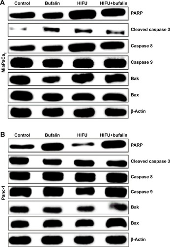
Figure 3 Bufalin enhances in vivo sensitivity of HIFU in PC xenograft models. MiaPaCa2 cells were injected into the right flank of BALB/C nude mice. After 14 days, the mice were randomized and treated with vehicle, HIFU, bufalin, and HIFU+bufalin for 21 days. The tumor growth in mice was monitored by caliper measurement every 3 days. (A) Tumor growth was shown as tumor volume. (B) Tumor weight in mice on Day 35 was compared in the four treatment groups. The data shown are representative of two independent experiments.
Notes: The error bars in (A) represent the SEM. N=6 mice for each group. *P<0.05, **P<0.01 and ***P<0.0001.
Abbreviations: HIFU, high-intensity focused ultrasound; ns, nonsignificant.
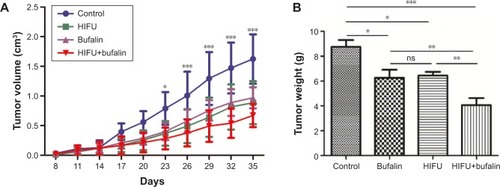
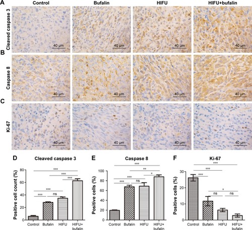
Figure 4 HIFU inhibits cell proliferation and increases cell death in MiaPaCa2 xenograft tumors. Tumors from mice treated with vehicle, bufalin, HIFU, or HIFU+bufalin for 35 days were subjected to immunohistochemical staining with cleaved caspase-3 (A and D), caspase-8 (B and E), and Ki-67 (C and F) antibodies.
Notes: All images are representative of three independent experiments. Scale bar=50 µm. *P<0.05, **P<0.01, and ***P<0.0001, one-way ANOVA. Abbreviations: HIFU, high-intensity focused ultrasound; ns, nonsignificant.