Figures & data
Figure 1 INPP4B promoted the proliferation of colon cancer cells by increasing mTORC1 activity. (A) The Western blotting analysis of pP70S6K-389, P70S6K, p4EBP1-65, 4EBP1, and INPP4B in control and SW620 cells with stable expression of INPP4B. Tubulin was used as a loading control. (B) The Western blotting analysis of pP70S6K-389, P70S6K, p4EBP1-65, 4EBP1, and INPP4B in HCT116 cells stably transduced with the control shRNA (shNC) or INPP4B shRNA (shINPP4B). Tubulin was used as a loading control. (C) Control and SW620 cells with a stable expression of INPP4B were seeded in 96-well plates. After 24 hours, cells were treated with DMSO or 0.1 μM of RAD001 for 48 hours. Then, cell proliferation was determined by CCK-8 assay. The results are presented as the mean ± SD of triplicate measurements. *P<0.05. (D) Control and SW620 cells with stable expression of INPP4B were treated with DMSO or 0.1 μM of RAD001. After 48 hours, cells were subjected to bromodeoxyuridine (BrdU) incorporation assays. The results are presented as the mean ± SD of triplicate measurements. *P<0.05.
Abbreviations: CCK-8, Cell Counting Kit-8; DMSO, dimethyl sulfoxide; BrdU, bromodeoxyuridine; shNC, control shRNA; NS, not significant.
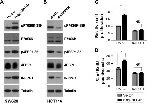
Figure 2 INPP4B activates cap-dependent translation in colon cancer cells. (A, B) Bicistronic luciferase assays. Control and SW620 cells with stable expression of INPP4B (A) or HCT116 cells with or without INPP4B depletion (B) were transfected with a bicistronic luciferase reporter plasmid. After 48 hours of transfection, luciferase activity was measured. Cap-dependent translational activity was determined, as described in the “Materials and methods” section. The results are presented as the mean ± SD of triplicate measurements. *P<0.05. (C, D) The m7GTP pull down assay. Cell lysates from control and SW620 cells with a stable expression of INPP4B (C), or HCT116 cells with or without INPP4B depletion (D) were precipitated with m7GTP sepharose beads and subjected to Western blot analysis with the indicated antibodies. (E) Control and SW620 cells with a stable expression of INPP4B transfected with the bicistronic luciferase reporter plasmid were treated with DMSO or 0.1 μM of RAD001 and subjected to luciferase activity measurement. The results are presented as the mean ± SD of triplicate measurements. *P<0.05.
Abbreviations: DMSO, dimethyl sulfoxide; shNC, control shRNA; NS, not significant.
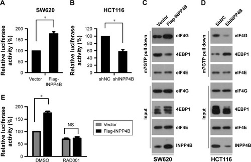
Figure 3 INPP4B promotes colon cancer cell proliferation by activating cap-dependent translation. (A) Control and SW620 cells with a stable expression of INPP4B were treated with DMSO or 40 μM 4EGI-1 for 48 hours, and subjected to Western blot analysis with the cyclinD1 antibody. Tubulin was used as a loading control. (B) Control and SW620 cells with a stable expression of INPP4B were treated with DMSO or 40 μM of 4EGI-1 for 48 hours, and harvested for real-time PCR analysis. The results are presented as the mean ± SD of triplicate measurements. (C) Control and SW620 cells with a stable expression of INPP4B were seeded in 96-well plates. After 24 hours, cells were treated with DMSO or 40 μM of 4EGI-1 for 48 hours. Then, cell proliferation was determined by CCK-8 assay. The results were presented as the mean ± SD of triplicate measurements. *P<0.05. (D) HCT116 cells with or without INPP4B depletion were transfected with the control siRNA (siNC) or 4EBP1 siRNA (si4EBP1). After 48 hours post-transfection, cells were harvested for Western blot analysis with the indicated antibodies. Tubulin was used as a loading control. (E) HCT116 cells with or without INPP4B depletion were co-transfected with siNC or si4EBP1 and a bicistronic luciferase reporter plasmid. At 48 hours after transfection, luciferase activity was measured. The results are presented as the mean ± SD of triplicate measurements. *P<0.05. (F) HCT116 cells with or without INPP4B depletion were transfected with siNC or si4EBP1. At 24 hours post-transfection, cells were seeded in 96-well plates. After 48 hours, cell proliferation was measured by CCK-8 assay. The results are presented as the mean ± SD of triplicate measurements. *P<0.05.
Abbreviations: DMSO, dimethyl sulfoxide; CCK-8, Cell Counting Kit-8; NS, not significant.
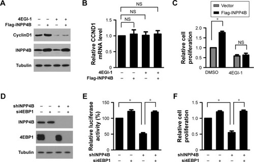
Figure 4 Akt and SGK3 regulate the cap-dependent translation downstream of INPP4B. (A) The Western blot analysis of AKT and SGK3 expression in control and SW620 cells with a stable expression of INPP4B co-introduced with AKT siRNA (siAKT) and SGK3 siRNA (siSGK3). Tubulin was used as a loading control. (B) Control and SW620 cells with a stable expression of INPP4B transfected with a bicistronic luciferase reporter plasmid were co-introduced with siAKT and siSGK3. After 48 hours of transfection, luciferase activity was measured. The results are presented as the mean ± SD of triplicate measurements. *P<0.05. (C) Control and SW620 cells with a stable expression of INPP4B were co-transfected with siAKT and siSGK3. At 24 hours post-transfection, cells were seeded in 96-well plates. After 48 hours, cell proliferation was measured by CCK-8 assay. The results are presented as the mean ± SD of triplicate measurements. *P<0.05. (D) Western blot analysis of AKT and SGK3 expression in HCT116 cells with or without INPP4B depletion co-introduced with myr-Akt and myr-SGK3 vectors. Tubulin was used as a loading control. (E) HCT116 cells with or without INPP4B depletion transfected with a bicistronic luciferase reporter plasmid were co-introduced with the myr-Akt and myr-SGK3 vectors. After 48 hours of transfection, luciferase activity was measured. The results are presented as the mean ± SD of triplicate measurements. *P<0.05. (F) HCT116 cells with or without INPP4B depletion, which were transfected with a bicistronic luciferase reporter plasmid, were co-introduced with the myr-Akt and myr-SGK3 vectors. At 24 hours post-transfection, cells were seeded in 96-well plates. After 48 hours, cell proliferation was measured by CCK-8 assay. The results are presented as the mean ± SD of triplicate measurements. *P<0.05.
Abbreviations: CCK-8, Cell Counting Kit-8; NS, not significant.
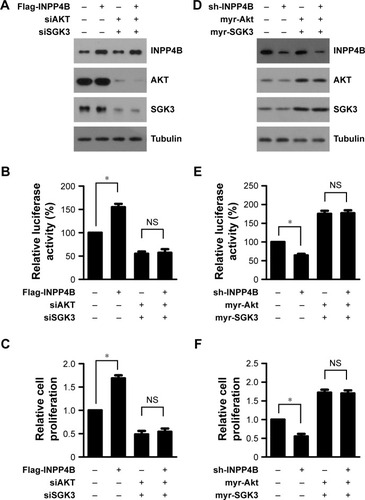
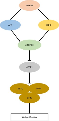
Figure 5 A working model for INPP4B function and mechanism in CRC cell proliferation.
Abbreviation: CRC, colorectal cancer.